


 癌症的基本特征包括细胞增殖、血管生成、迁移、凋亡逃避机制和细胞永生等。找到癌症发生过程中这些通路的关键标记物和对应的抗体用于检测至关重要。
 为您推荐一个泛素化位点预测神器——泛素化分析工具,可以为您的蛋白的泛素化位点作出预测和评分。
 细胞自噬受体图形绘图工具为你的蛋白的细胞受体结合位点作出预测和评分,识别结合到自噬通路中的蛋白是非常重要的,便于让我们理解自噬在正常生理、病理过程中的作用,如发育、细胞分化、神经退化性疾病、压力条件下、感染和癌症。
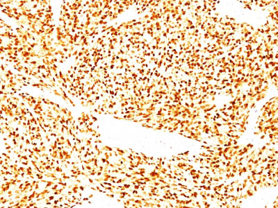
MyoD1 (Rhabdomyosarcoma Marker) Antibody - With BSA and Azide
Mouse Monoclonal Antibody [Clone SPM427 ]
- 产品详情
- 实验流程
- 背景知识
Application
| IF, FC, IHC-P |
|---|---|
| Primary Accession | P15172 |
| Other Accession | 4654, 181768 |
| Reactivity | Human, Mouse, Rat, Chicken |
| Host | Mouse |
| Clonality | Monoclonal |
| Isotype | Mouse / IgG1, kappa |
| Clone Names | SPM427 |
| Calculated MW | 34501 Da |
| Gene ID | 4654 |
|---|---|
| Other Names | Myoblast determination protein 1, Class C basic helix-loop-helix protein 1, bHLHc1, Myogenic factor 3, Myf-3, MYOD1, BHLHC1, MYF3, MYOD |
| Application Note | IF~~1:50~200 FC~~1:10~50 IHC-P~~1:50~200 |
| Format | 200ug/ml of Ab purified from Bioreactor Concentrate by Protein A/G. Prepared in 10mM PBS with 0.05% BSA & 0.05% azide. Also available WITHOUT BSA at 1.0mg/ml. |
| Storage | Store at 2 to 8°C.Antibody is stable for 24 months. |
| Precautions | MyoD1 (Rhabdomyosarcoma Marker) Antibody - With BSA and Azide is for research use only and not for use in diagnostic or therapeutic procedures. |
| Name | MYOD1 |
|---|---|
| Synonyms | BHLHC1, MYF3, MYOD |
| Function | Acts as a transcriptional activator that promotes transcription of muscle-specific target genes and plays a role in muscle differentiation. Together with MYF5 and MYOG, co-occupies muscle-specific gene promoter core region during myogenesis. Induces fibroblasts to differentiate into myoblasts. Interacts with and is inhibited by the twist protein. This interaction probably involves the basic domains of both proteins (By similarity). |
| Cellular Location | Nucleus. |
For Research Use Only. Not For Use In Diagnostic Procedures.
Provided below are standard protocols that you may find useful for product applications.
BACKGROUND
Recognizes a phosphor-protein of 45kDa, identified as MyoD1. The epitope of this MAb maps between amino acid 180-189 in the C-terminal of mouse MyoD1 protein. It does not cross react with myogenin, Myf5, or Myf6. Antibody to MyoD1 labels the nuclei of myoblasts in developing muscle tissues. MyoD1 is not detected in normal adult tissue, but is highly expressed in the tumor cell nuclei of rhabdomyosarcomas. Occasionally nuclear expression of MyoD1 is seen in ectomesenchymoma and a subset of Wilmā€™s tumors. Weak cytoplasmic staining is observed in several non-muscle tissues, including glandular epithelium and also in rhabdomyosarcomas, neuroblastomas, Ewingā€™s sarcomas and alveolar soft part sarcomas.
REFERENCES
Thulasi R et. al. Cell Growth and Differentiation, 1996, 7(4):531-41. | Wesche WA et. al. American Journal of Surgical Pathology, 1995, 19(3):261-9. | Parham DM et. al. Acta Neuropathologica, 1994, 87:605-11
终于等到您。ABCEPTA(百远生物)抗体产品。
点击下方“我要评价 ”按钮提交您的反馈信息,您的反馈和评价是我们最宝贵的财富之一,
我们将在1-3个工作日内处理您的反馈信息。
如有疑问,联系:0512-88856768 tech-china@abcepta.com.


