


 癌症的基本特征包括细胞增殖、血管生成、迁移、凋亡逃避机制和细胞永生等。找到癌症发生过程中这些通路的关键标记物和对应的抗体用于检测至关重要。
 为您推荐一个泛素化位点预测神器——泛素化分析工具,可以为您的蛋白的泛素化位点作出预测和评分。
 细胞自噬受体图形绘图工具为你的蛋白的细胞受体结合位点作出预测和评分,识别结合到自噬通路中的蛋白是非常重要的,便于让我们理解自噬在正常生理、病理过程中的作用,如发育、细胞分化、神经退化性疾病、压力条件下、感染和癌症。
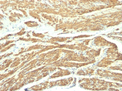
Caldesmon, HMW (h-Caldesmon) (Smooth Muscle Marker) Antibody - With BSA and Azide
Mouse Monoclonal Antibody [Clone SPM168 ]
- 产品详情
- 实验流程
- 背景知识
Application
| IHC, IF, FC |
|---|---|
| Primary Accession | Q05682 |
| Other Accession | 800, 490203 |
| Reactivity | Human |
| Host | Mouse |
| Clonality | Monoclonal |
| Isotype | Mouse / IgG1, kappa |
| Clone Names | SPM168 |
| Calculated MW | 93231 Da |
| Gene ID | 800 |
|---|---|
| Other Names | Caldesmon, CDM, CALD1, CAD, CDM |
| Application Note | IHC~~1:100~500 IF~~1:50~200 FC~~1:10~50 |
| Storage | Store at 2 to 8°C.Antibody is stable for 24 months. |
| Precautions | Caldesmon, HMW (h-Caldesmon) (Smooth Muscle Marker) Antibody - With BSA and Azide is for research use only and not for use in diagnostic or therapeutic procedures. |
| Name | CALD1 |
|---|---|
| Synonyms | CAD, CDM |
| Function | Actin- and myosin-binding protein implicated in the regulation of actomyosin interactions in smooth muscle and nonmuscle cells (could act as a bridge between myosin and actin filaments). Stimulates actin binding of tropomyosin which increases the stabilization of actin filament structure. In muscle tissues, inhibits the actomyosin ATPase by binding to F-actin. This inhibition is attenuated by calcium-calmodulin and is potentiated by tropomyosin. Interacts with actin, myosin, two molecules of tropomyosin and with calmodulin. Also plays an essential role during cellular mitosis and receptor capping. Involved in Schwann cell migration during peripheral nerve regeneration (By similarity). |
| Cellular Location | Cytoplasm, cytoskeleton {ECO:0000250|UniProtKB:P13505}. Cytoplasm, myofibril {ECO:0000250|UniProtKB:P13505}. Cytoplasm, cytoskeleton, stress fiber {ECO:0000250|UniProtKB:P13505}. Note=On thin filaments in smooth muscle and on stress fibers in fibroblasts (nonmuscle) {ECO:0000250|UniProtKB:P13505} |
| Tissue Location | High-molecular-weight caldesmon (isoform 1) is predominantly expressed in smooth muscles, whereas low-molecular-weight caldesmon (isoforms 2, 3, 4 and 5) are widely distributed in non-muscle tissues and cells. Not expressed in skeletal muscle or heart |
For Research Use Only. Not For Use In Diagnostic Procedures.
Provided below are standard protocols that you may find useful for product applications.
BACKGROUND
Recognizes a protein of 150kDa, which is identified as the high molecular weight variant of Caldesmon. Two closely related variants of human caldesmon have been identified which are different in their electrophoretic mobility and cellular distribution. The h-caldesmon variant (120-150kDa) is predominantly expressed in smooth muscle whereas l-caldesmon (70-80kDa) is found in non- muscle tissue and cells. Neither of the two variants has been detected in skeletal muscle. This MAb recognizes only the 150kDa variant (h-caldesmon) in Western blots of human aortic media extracts and is unreactive with fibroblast extracts from cultivated human foreskin. Caldesmon is a developmentally regulated protein involved in smooth muscle and non-muscle contraction.
REFERENCES
Frid MG, et al. Phenotypic changes of human smooth muscle cells during development: Late expression of heavy caldesmon and calponin. Dev Biol 1992; 153:185 |
终于等到您。ABCEPTA(百远生物)抗体产品。
点击下方“我要评价 ”按钮提交您的反馈信息,您的反馈和评价是我们最宝贵的财富之一,
我们将在1-3个工作日内处理您的反馈信息。
如有疑问,联系:0512-88856768 tech-china@abcepta.com.


