


 癌症的基本特征包括细胞增殖、血管生成、迁移、凋亡逃避机制和细胞永生等。找到癌症发生过程中这些通路的关键标记物和对应的抗体用于检测至关重要。
 为您推荐一个泛素化位点预测神器——泛素化分析工具,可以为您的蛋白的泛素化位点作出预测和评分。
 细胞自噬受体图形绘图工具为你的蛋白的细胞受体结合位点作出预测和评分,识别结合到自噬通路中的蛋白是非常重要的,便于让我们理解自噬在正常生理、病理过程中的作用,如发育、细胞分化、神经退化性疾病、压力条件下、感染和癌症。
PNL2 (Melanoma Associated Antigen) Antibody - With BSA and Azide
Mouse Monoclonal Antibody [Clone PNL2 ]
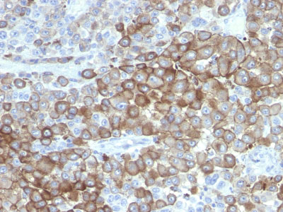
- 产品详情
- 实验流程
- 背景知识
Application 
| IHC, IF |
|---|---|
| Reactivity | Human, Mouse, Drosophila |
| Host | Mouse |
| Clonality | Monoclonal |
| Isotype | Mouse / IgG1 |
| Clone Names | PNL2 |
| Calculated MW | 160 KDa |
| Application Note | IHC~~1:100~500 IF~~1:50~200 |
|---|---|
| Storage | Store at 2 to 8°C.Antibody is stable for 24 months. |
| Precautions | PNL2 (Melanoma Associated Antigen) Antibody - With BSA and Azide is for research use only and not for use in diagnostic or therapeutic procedures. |
For Research Use Only. Not For Use In Diagnostic Procedures.
Provided below are standard protocols that you may find useful for product applications.
BACKGROUND
Anti-PNL2 is a novel monoclonal antibody, which has recently been introduced as an immunohistochemical reagent to stain melanocytes and tumors derived therefrom. The antigen recognized by PNL2 is different from Melan A and gp100. Its epitope is not destroyed by digestion with neuraminidase i.e. its epitope id not glycosylated. Anti-PNL2 may be most useful because of its high sensitivity for metastatic melanoma (87%), as opposed to 76% for anti-HMB45 and 82% for anti-MART-1. Anti-PNL2 labels intra-epidermal nevi while the dermal component of compound nevi are largely non-reactive with anti-PNL2. Antibodies against PNL2, MART-1 (Melan A) and HMB45 stain most clear cell sarcoma cells and a few cells in angio-myolipomas and lymphangioleiomyomatosis. Anti-PNL2 is a useful antibody for the identification of melanomas and clear cell sarcomas. Differential diagnosis is aided by the results from a panel of antibodies, including antibodies against HMB45, MART-1, tyrosinase, and MiTF.
REFERENCES
Rochaix, P et al. PNL2, a New Monoclonal Antibody Directed against a Fixative-Resistant Melanocyte Antigen. Mod Pathol 2003;16(5):481-490. | J. A. Ramos-Vara and M. A. Miller. Immunohistochemical Identification of Canine Melanocytic Neoplasms With Antibodies to Melanocytic Antigen PNL2 and Tyrosinase: Comparison With Melan A. Veterinary Pathology OnlineFirst, published on September 21, 2010 as doi:10.1177/0300985810382095

终于等到您。ABCEPTA(百远生物)抗体产品。
点击下方“我要评价 ”按钮提交您的反馈信息,您的反馈和评价是我们最宝贵的财富之一,
我们将在1-3个工作日内处理您的反馈信息。
如有疑问,联系:0512-88856768 tech-china@abcepta.com.


