


 癌症的基本特征包括细胞增殖、血管生成、迁移、凋亡逃避机制和细胞永生等。找到癌症发生过程中这些通路的关键标记物和对应的抗体用于检测至关重要。
 为您推荐一个泛素化位点预测神器——泛素化分析工具,可以为您的蛋白的泛素化位点作出预测和评分。
 细胞自噬受体图形绘图工具为你的蛋白的细胞受体结合位点作出预测和评分,识别结合到自噬通路中的蛋白是非常重要的,便于让我们理解自噬在正常生理、病理过程中的作用,如发育、细胞分化、神经退化性疾病、压力条件下、感染和癌症。
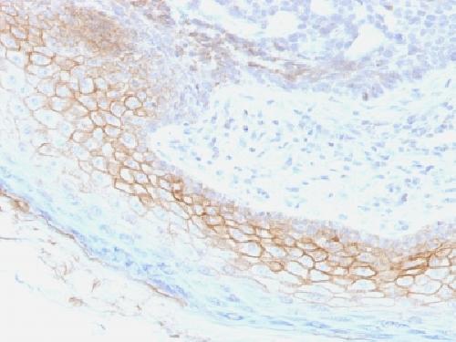
Anti-Desmocollin-2/3 Antibody
Mouse Monoclonal Antibody
- 产品详情
- 实验流程
- 背景知识
Application
| WB, IHC-P, IF, FC |
|---|---|
| Primary Accession | Q02487 |
| Other Accession | 95612 |
| Reactivity | Human, Mouse, Rat |
| Host | Mouse |
| Clonality | Monoclonal |
| Isotype | Mouse / IgG1, kappa |
| Clone Names | 7G6 |
| Calculated MW | 99962 Da |
| Gene ID | 1824 |
|---|---|
| Other Names | ARVD11; Cadherin family member 2; CDHF2; Desmocollin-2; Desmocollin-3; Desmosomal glycoprotein II and III; Desmosomal glycoprotein II; Desmosomal glycoprotein III; DG2; DGII/III; DSC2; DSC3 |
| Application Note | Flow Cytometry (0.5-1ug/million cells); Immunofluorescence (1-2ug/ml); ,Western Blotting (0.5-1.0ug/ml); ,Immunohistology (Formalin-fixed) (1-2ug/ml for 30 minutes at RT),(Staining of formalin-fixed tissues requires boiling tissue sections in 10mM Tris with 1mM EDTA, pH 9.0, for 10-20 min followed by cooling at RT for 20 minutes),Optimal dilution for a specific application should be determined. |
| Format | 200ug/ml of Ab purified from Bioreactor Concentrate by Protein A/G. Prepared in 10mM PBS with 0.05% BSA & 0.05% azide. Also available WITHOUT BSA & azide at 1.0mg/ml. |
| Storage | Store at 2 to 8°C.Antibody is stable for 24 months. |
| Precautions | Anti-Desmocollin-2/3 Antibody is for research use only and not for use in diagnostic or therapeutic procedures. |
| Name | DSC2 (HGNC:3036) |
|---|---|
| Synonyms | CDHF2, DSC3 |
| Function | A component of desmosome cell-cell junctions which are required for positive regulation of cellular adhesion (PubMed:33596089). Promotes timely incorporation of DSG2 into desmosome intercellular junctions and promotes interaction of desmosome cell junctions with intermediate filament cytokeratin, via modulation of DSP phosphorylation (PubMed:33596089). Plays an important role in desmosome-mediated maintenance of intestinal epithelial cell intercellular adhesion strength and barrier function (PubMed:33596089). Positively regulates wound healing of intestinal mucosa via promotion of epithelial cell migration, and also plays a role in mechanotransduction of force between intestinal epithelial cells and extracellular matrix (PubMed:31967937). May contribute to epidermal cell positioning (stratification) by mediating differential adhesiveness between cells that express different isoforms. May promote p38MAPK signaling activation that facilitates keratinocyte migration (By similarity). |
| Cellular Location | Cell membrane; Single-pass type I membrane protein. Cell junction, desmosome |
| Tissue Location | Expressed at intercalated disks in the heart, where it is colocalized with CDH2 (at protein level) (PubMed:23863954, PubMed:33784018). Expressed in intestinal mucosal cells (at protein level) (PubMed:31967937). |
For Research Use Only. Not For Use In Diagnostic Procedures.
Provided below are standard protocols that you may find useful for product applications.
BACKGROUND
Desmosomes are intercellular adhering junctions that represent cell surface attachment sites for intermediate filament. The desmosome is subdivided into two regions. The plaque region lies adjacent to the plasma, and is believed to contain molecules that attach the intermediate filament cytoskeleton to the desmosome. The core region is composed of transmembrane glycoproteins that are thought to mediate cell-cell adhesion. Desmogleins and desmocollins are the main desmosomal transmembrane proteins. These desmosomal glycoproteins belong to the members of the cadherin family of adhesion molecules. Three different isoforms of both desmogleins and desmocollins have been identified, named as desmoglein 1-3 and desmocollins. Desmosomal cadherins showed differentiation-specific expression in the human epidermis, although the functional significance of this differential expression is not fully understood. Desmocollin-1 can be found in the upper layers. The expression of desmocollin-2 varies in the basal and suprabasal layers. And desmocollin-3 is expressed more evenly throughout the suprabasal layers.
终于等到您。ABCEPTA(百远生物)抗体产品。
点击下方“我要评价 ”按钮提交您的反馈信息,您的反馈和评价是我们最宝贵的财富之一,
我们将在1-3个工作日内处理您的反馈信息。
如有疑问,联系:0512-88856768 tech-china@abcepta.com.


