


 癌症的基本特征包括细胞增殖、血管生成、迁移、凋亡逃避机制和细胞永生等。找到癌症发生过程中这些通路的关键标记物和对应的抗体用于检测至关重要。
 为您推荐一个泛素化位点预测神器——泛素化分析工具,可以为您的蛋白的泛素化位点作出预测和评分。
 细胞自噬受体图形绘图工具为你的蛋白的细胞受体结合位点作出预测和评分,识别结合到自噬通路中的蛋白是非常重要的,便于让我们理解自噬在正常生理、病理过程中的作用,如发育、细胞分化、神经退化性疾病、压力条件下、感染和癌症。
Anti-ALDH1A1 (Aldehyde Dehydrogenase 1A1) Antibody
Mouse Monoclonal Antibody
- 产品详情
- 实验流程
- 背景知识
Application
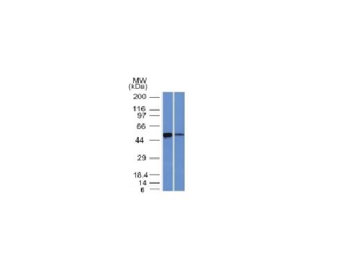
| WB, IHC-P, IF, FC |
|---|---|
| Primary Accession | P00352 |
| Other Accession | 76392 |
| Reactivity | Human |
| Host | Mouse |
| Clonality | Monoclonal |
| Isotype | Mouse / IgG1 |
| Clone Names | ALDH1A1/1381 |
| Calculated MW | 54862 Da |
| Gene ID | 216 |
|---|---|
| Other Names | Acetaldehyde dehydrogenase 1; AHD2; ALDC; Aldehyde dehydrogenase 1 soluble; Aldehyde dehydrogenase 1A1; Aldehyde dehydrogenase family 1 member A1; ALDH-E1; ALDH1; ALDH1A1; epididymis luminal protein 12; epididymis luminal protein 9; epididymis secretory sperm binding protein Li 53e; HEL-S-53e; PUMB1; RALDH1; Retinal dehydrogenase 1 |
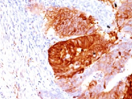
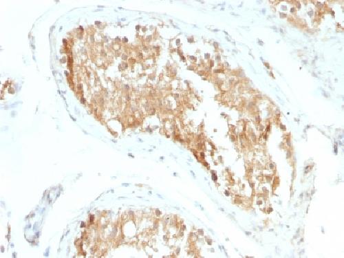
| Application Note | Flow Cytometry (0.5-1ug/million cells); Immunofluorescence (1-2ug/ml); ,Western Blotting (0.5-1ug/ml); ,Immunohistology (Formalin-fixed) (0.5-1ug/ml for 30 min at RT),(Staining of formalin-fixed tissues requires boiling tissue sections in 10mM Citrate Buffer, pH 6.0, for 10-20 min followed by cooling at RT for 20 minutes),Optimal dilution for a specific application should be determined. |
| Format | 200ug/ml of Ab purified from Bioreactor Concentrate by Protein A/G. Prepared in 10mM PBS with 0.05% BSA & 0.05% azide. Also available WITHOUT BSA & azide at 1.0mg/ml. |
| Storage | Store at 2 to 8°C.Antibody is stable for 24 months. |
| Precautions | Anti-ALDH1A1 (Aldehyde Dehydrogenase 1A1) Antibody is for research use only and not for use in diagnostic or therapeutic procedures. |
| Name | ALDH1A1 (HGNC:402) |
|---|---|
| Function | Cytosolic dehydrogenase that catalyzes the irreversible oxidation of a wide range of aldehydes to their corresponding carboxylic acid (PubMed:12941160, PubMed:15623782, PubMed:17175089, PubMed:19296407, PubMed:25450233, PubMed:26373694). Functions downstream of retinol dehydrogenases and catalyzes the oxidation of retinaldehyde into retinoic acid, the second step in the oxidation of retinol/vitamin A into retinoic acid (By similarity). This pathway is crucial to control the levels of retinol and retinoic acid, two important molecules which excess can be teratogenic and cytotoxic (By similarity). Also oxidizes aldehydes resulting from lipid peroxidation like (E)-4-hydroxynon-2-enal/HNE, malonaldehyde and hexanal that form protein adducts and are highly cytotoxic. By participating for instance to the clearance of (E)-4-hydroxynon-2-enal/HNE in the lens epithelium prevents the formation of HNE-protein adducts and lens opacification (PubMed:12941160, PubMed:15623782, PubMed:19296407). Also functions downstream of fructosamine-3-kinase in the fructosamine degradation pathway by catalyzing the oxidation of 3-deoxyglucosone, the carbohydrate product of fructosamine 3-phosphate decomposition, which is itself a potent glycating agent that may react with lysine and arginine side-chains of proteins (PubMed:17175089). Also has an aminobutyraldehyde dehydrogenase activity and is probably part of an alternative pathway for the biosynthesis of GABA/4-aminobutanoate in midbrain, thereby playing a role in GABAergic synaptic transmission (By similarity). |
| Cellular Location | Cytoplasm, cytosol. Cell projection, axon {ECO:0000250|UniProtKB:P24549} |
| Tissue Location | Expressed by erythrocytes (at protein level). |
For Research Use Only. Not For Use In Diagnostic Procedures.
Provided below are standard protocols that you may find useful for product applications.
BACKGROUND
ALDH1A1 belongs to the ALDH enzymes, a family of evolutionarily conserved enzymes comprised of 19 isoforms that are localized in the cytoplasm, mitochondria or nucleus. ALDH1A1 is predominantly expressed in the epithelium of testis, brain, eye, liver, kidney, as well as neural and hematopoietic stem cells. Reportedly, high ALDH1A1 expression is found in solitary fibrous tumor (SFT) and hemangiopericytoma (HPC), compared to meningiomas and synovial sarcomas. In combination with CD34, ALDH1A1 may be useful for the differentiation among SFT, HPC, meningioma, and synovial sarcoma.
终于等到您。ABCEPTA(百远生物)抗体产品。
点击下方“我要评价 ”按钮提交您的反馈信息,您的反馈和评价是我们最宝贵的财富之一,
我们将在1-3个工作日内处理您的反馈信息。
如有疑问,联系:0512-88856768 tech-china@abcepta.com.


