


 癌症的基本特征包括细胞增殖、血管生成、迁移、凋亡逃避机制和细胞永生等。找到癌症发生过程中这些通路的关键标记物和对应的抗体用于检测至关重要。
 为您推荐一个泛素化位点预测神器——泛素化分析工具,可以为您的蛋白的泛素化位点作出预测和评分。
 细胞自噬受体图形绘图工具为你的蛋白的细胞受体结合位点作出预测和评分,识别结合到自噬通路中的蛋白是非常重要的,便于让我们理解自噬在正常生理、病理过程中的作用,如发育、细胞分化、神经退化性疾病、压力条件下、感染和癌症。
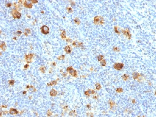
Anti-CD15 / FUT4 (Reed-Sternberg Cell Marker) Antibody
Recombinant Rabbit Monoclonal Antibody
- 产品详情
- 实验流程
- 背景知识
Application
| IHC-P, IF, FC |
|---|---|
| Primary Accession | P22083 |
| Other Accession | 654379 |
| Reactivity | Human, Mouse, Rat |
| Host | Rabbit |
| Clonality | Monoclonal |
| Isotype | Rabbit / IgG, kappa |
| Clone Names | FUT4/1478R |
| Calculated MW | 59084 Da |
| Gene ID | 2526 |
|---|---|
| Other Names | 3 Fucosyl N Acetyl Lactosamine; Alpha (1, 3) Fucosyltransferase; Alpha 13 fucosyltransferase FucT; ELAM Ligand Fucosyltransferase; ELFT; FCT3A; Fuc-TIV; Fucosyltransferase 4 Alpha 1 3 Fucosyltransferase Myeloid Specific; Fucosyltransferase 4; Galactoside 3 L Fucosyltransferase; Lewis X; LeX; SSEA1; Stage Specific Embryonic Antigen 1 |
| Application Note | Flow Cytometry (2-4ug/million cells); Immunofluorescence (2-4ug/ml); ,Immunohistology (Formalin-fixed) (5-10ug/ml for 30 minutes at RT),(Staining of formalin-fixed tissues requires boiling tissue sections in 10mM Tris with 1mM EDTA, pH 9.0, for 10-20 min followed by cooling at RT for 20 minutes),Optimal dilution for a specific application should be determined. |
| Format | 200ug/ml of Ab purified by Protein A Column. Prepared in 10mM PBS with 0.05% BSA & 0.05% azide. Also available WITHOUT BSA & Azide at 1.0mg/ml. |
| Storage | Store at 2 to 8°C.Antibody is stable for 24 months. |
| Precautions | Anti-CD15 / FUT4 (Reed-Sternberg Cell Marker) Antibody is for research use only and not for use in diagnostic or therapeutic procedures. |
| Name | FUT4 {ECO:0000303|PubMed:29593094} |
|---|---|
| Function | [Isoform Short]: Catalyzes alpha(1->3) linkage of fucosyl moiety transferred from GDP-beta-L-fucose to N-acetyl glucosamine (GlcNAc) within type 2 lactosamine (LacNAc, Gal-beta(1->4)GlcNAc) glycan attached to N- or O-linked glycoproteins (PubMed:1702034, PubMed:1716630, PubMed:29593094). Robustly fucosylates nonsialylated distal LacNAc unit of the polylactosamine chain to form Lewis X antigen (CD15), a glycan determinant known to mediate important cellular functions in development and immunity. Fucosylates with lower efficiency sialylated LacNAc acceptors to form sialyl Lewis X and 6- sulfo sialyl Lewis X determinants that serve as recognition epitopes for C-type lectins (PubMed:1716630, PubMed:29593094). Together with FUT7 contributes to SELE, SELL and SELP selectin ligand biosynthesis and selectin-dependent lymphocyte homing, leukocyte migration and blood leukocyte homeostasis (By similarity). In a cell type specific manner, may also fucosylate the internal LacNAc unit of the polylactosamine chain to form VIM-2 antigen that serves as recognition epitope for SELE (PubMed:11278338, PubMed:1716630). |
| Cellular Location | Golgi apparatus, Golgi stack membrane; Single- pass type II membrane protein. Note=Membrane-bound form in trans cisternae of Golgi |
| Tissue Location | [Isoform Short]: Expressed at low levels in bone marrow-derived mesenchymal stem cells. |
For Research Use Only. Not For Use In Diagnostic Procedures.
Provided below are standard protocols that you may find useful for product applications.
BACKGROUND
CD15 plays a role in mediating phagocytosis, bactericidal activity, and chemotaxis. It is present on >95% of granulocytes including neutrophils and eosinophils and to a lesser degree on monocytes. In addition, CD15 is expressed in Reed-Sternberg cells and some epithelial cells. CD15 antibody is very useful in the identification of Hodgkin s disease. CD15 is occasionally expressed in large cell lymphomas of both B and T phenotypes which otherwise have a quite distinct histological appearance.
终于等到您。ABCEPTA(百远生物)抗体产品。
点击下方“我要评价 ”按钮提交您的反馈信息,您的反馈和评价是我们最宝贵的财富之一,
我们将在1-3个工作日内处理您的反馈信息。
如有疑问,联系:0512-88856768 tech-china@abcepta.com.


