


 癌症的基本特征包括细胞增殖、血管生成、迁移、凋亡逃避机制和细胞永生等。找到癌症发生过程中这些通路的关键标记物和对应的抗体用于检测至关重要。
 为您推荐一个泛素化位点预测神器——泛素化分析工具,可以为您的蛋白的泛素化位点作出预测和评分。
 细胞自噬受体图形绘图工具为你的蛋白的细胞受体结合位点作出预测和评分,识别结合到自噬通路中的蛋白是非常重要的,便于让我们理解自噬在正常生理、病理过程中的作用,如发育、细胞分化、神经退化性疾病、压力条件下、感染和癌症。
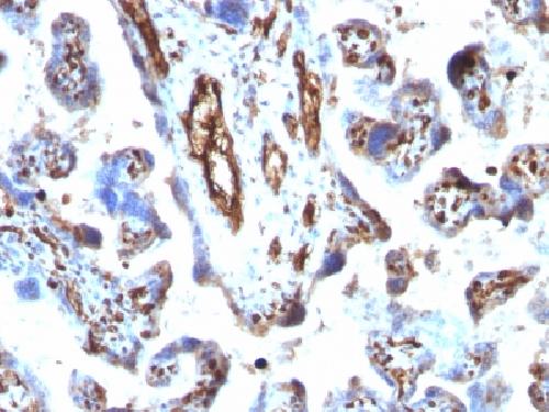
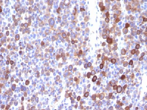
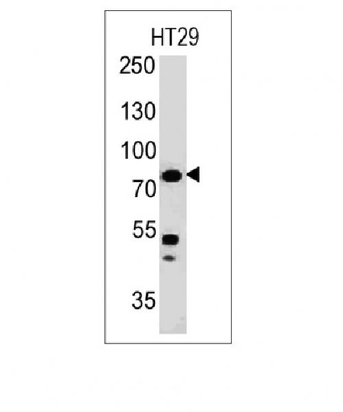
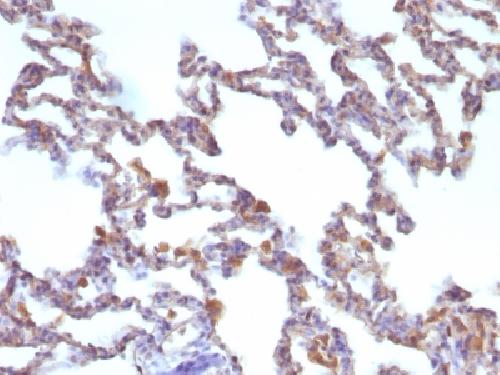
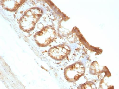
Anti-Moesin Antibody
Mouse Monoclonal Antibody
- 产品详情
- 实验流程
- 背景知识
Application
| WB, IHC-P, IF, FC |
|---|---|
| Primary Accession | P26038 |
| Other Accession | 87752 |
| Reactivity | Human, Rat |
| Host | Mouse |
| Clonality | Monoclonal |
| Isotype | Mouse / IgG1, kappa |
| Clone Names | MSN/491 |
| Other Names | Membrane-organizing extension spike protein; Moesin/anaplastic lymphoma kinase fusion protein; MSN/ALK fusion |
|---|---|
| Application Note | Flow Cytometry (0.5-1ug/million cells); Immunofluorescence (0.5-1ug/ml); ,Western Blotting (0.5-1ug/ml); ,Immunohistology (Formalin-fixed) (0.5-1ug/ml for 30 minutes at RT),(Staining of formalin-fixed tissues requires boiling tissue sections in 10mM citrate buffer, pH 6.0, for 10-20 min followed by cooling at RT for 20 minutes),Optimal dilution for a specific application should be determined. |
| Format | 200ug/ml of Ab purified from Bioreactor Concentrate by Protein A/G. Prepared in 10mM PBS with 0.05% BSA & 0.05% azide. Also available WITHOUT BSA & azide at 1.0mg/ml. |
| Storage | Store at 2 to 8°C.Antibody is stable for 24 months. |
| Precautions | Anti-Moesin Antibody is for research use only and not for use in diagnostic or therapeutic procedures. |
For Research Use Only. Not For Use In Diagnostic Procedures.
Provided below are standard protocols that you may find useful for product applications.
BACKGROUND
Recognizes 78kDa moesin protein. Moesin, a member of the talin-4.1 superfamily, is a linking protein of the sub-membranous actin cytoskeleton. It is expressed in variable amounts in cells of different phenotypes such as macrophages, lymphocytes, fibroblastic, endothelial, epithelial, and neuronal cell lines but not in blood cells. The ERM proteins, ezrin, radixin, and moesin are involved in a variety of cellular functions, such as cell adhesion, migration, and the organization of cell surface structures, and are highly homologous, both in protein sequence and in functional activity, with merlin/schwannomin, a neurofibromatosis-2-associated tumor-suppressor protein. Cell lines of epithelial and mesothelial origin contain both moesin and radixin whereas cells of endothelial and lymphoid origin express moesin.
终于等到您。ABCEPTA(百远生物)抗体产品。
点击下方“我要评价 ”按钮提交您的反馈信息,您的反馈和评价是我们最宝贵的财富之一,
我们将在1-3个工作日内处理您的反馈信息。
如有疑问,联系:0512-88856768 tech-china@abcepta.com.


