


 癌症的基本特征包括细胞增殖、血管生成、迁移、凋亡逃避机制和细胞永生等。找到癌症发生过程中这些通路的关键标记物和对应的抗体用于检测至关重要。
 为您推荐一个泛素化位点预测神器——泛素化分析工具,可以为您的蛋白的泛素化位点作出预测和评分。
 细胞自噬受体图形绘图工具为你的蛋白的细胞受体结合位点作出预测和评分,识别结合到自噬通路中的蛋白是非常重要的,便于让我们理解自噬在正常生理、病理过程中的作用,如发育、细胞分化、神经退化性疾病、压力条件下、感染和癌症。
Anti-PDCD1 / PD1 / CD279 (Programmed Cell Death 1) Antibody
Recombinant Rabbit Monoclonal Antibody
- 产品详情
- 实验流程
- 背景知识
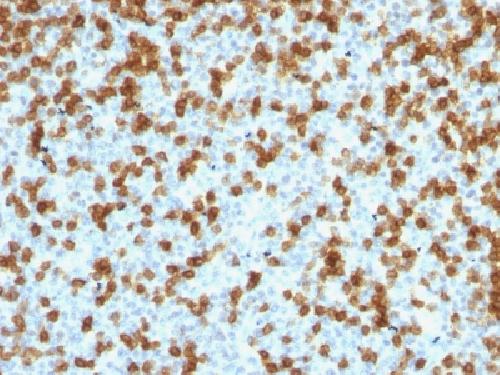
Application
| IHC-P, IF, FC |
|---|---|
| Primary Accession | Q15116 |
| Other Accession | 158297 |
| Reactivity | Human |
| Host | Rabbit |
| Clonality | Monoclonal |
| Isotype | Rabbit / IgG |
| Clone Names | PDCD1/1410R |
| Calculated MW | 31647 Da |
| Gene ID | 5133 |
|---|---|
| Other Names | CD279; hPD-1; hSLE1; PD1; PDCD1; Programmed Cell Death Protein 1; Protein PD-1; SLEB2; Systemic lupus erythematosus susceptibility 2 |
| Application Note | Flow Cytometry (0.5-1ug/million cells); Immunofluorescence (1-2ug/ml); ,Immunohistology (Formalin-fixed) (1-2ug/ml for 30 minutes at RT),(Staining of formalin-fixed tissues requires boiling tissue sections in 10mM Tris with 1mM EDTA, pH 9.0, for 10-20 min followed by cooling at RT for 20 minutes),Optimal dilution for a specific application should be determined. |
| Format | 200ug/ml of Ab purified by Protein A/G. Prepared in 10mM PBS with 0.05% BSA & 0.05% azide. Also available WITHOUT BSA & azide at 1.0mg/ml. |
| Storage | Store at 2 to 8°C.Antibody is stable for 24 months. |
| Precautions | Anti-PDCD1 / PD1 / CD279 (Programmed Cell Death 1) Antibody is for research use only and not for use in diagnostic or therapeutic procedures. |
| Name | PDCD1 {ECO:0000303|PubMed:7851902, ECO:0000312|HGNC:HGNC:8760} |
|---|---|
| Function | Inhibitory receptor on antigen activated T-cells that plays a critical role in induction and maintenance of immune tolerance to self (PubMed:21276005, PubMed:31754127, PubMed:32184441, PubMed:37208329). Delivers inhibitory signals upon binding to ligands CD274/PDCD1L1 and CD273/PDCD1LG2 (PubMed:21276005, PubMed:26602187). Following T-cell receptor (TCR) engagement, PDCD1 associates with TCR-CD3 in the immunological synapse and directly inhibits T-cell activation (PubMed:32184441). Suppresses T-cell activation through the recruitment of PTPN11/SHP-2: following ligand-binding, PDCD1 is phosphorylated within the ITSM motif, leading to the recruitment of the protein tyrosine phosphatase PTPN11/SHP-2 that mediates dephosphorylation of key TCR proximal signaling molecules, such as ZAP70, PRKCQ/PKCtheta and CD247/CD3zeta (PubMed:32184441). |
| Cellular Location | Cell membrane; Single-pass type I membrane protein |
For Research Use Only. Not For Use In Diagnostic Procedures.
Provided below are standard protocols that you may find useful for product applications.
BACKGROUND
PDCD-1 (programmed cell death-1 protein), also designated CD279, is a type I transmembrane receptor and a member of the immunoglobin gene superfamily. It is expressed on activated T-cells, B-cells, and myeloid cells. Anti-PDCD-1 is a marker of angioimmunoblastic lymphoma and suggests a unique cell of origin for this neoplasm. Unlike CD10 and BCL6, PDCD-1 is expressed by few B-cells, so anti-PDCD-1 may be a more specific and useful diagnostic marker in angioimmunoblastic lymphoma. In addition, PDCD-1 expression provides evidence that angioimmunoblastic lymphoma is a neoplasm derived from germinal center-associated T-cells.
终于等到您。ABCEPTA(百远生物)抗体产品。
点击下方“我要评价 ”按钮提交您的反馈信息,您的反馈和评价是我们最宝贵的财富之一,
我们将在1-3个工作日内处理您的反馈信息。
如有疑问,联系:0512-88856768 tech-china@abcepta.com.


