


 癌症的基本特征包括细胞增殖、血管生成、迁移、凋亡逃避机制和细胞永生等。找到癌症发生过程中这些通路的关键标记物和对应的抗体用于检测至关重要。
 为您推荐一个泛素化位点预测神器——泛素化分析工具,可以为您的蛋白的泛素化位点作出预测和评分。
 细胞自噬受体图形绘图工具为你的蛋白的细胞受体结合位点作出预测和评分,识别结合到自噬通路中的蛋白是非常重要的,便于让我们理解自噬在正常生理、病理过程中的作用,如发育、细胞分化、神经退化性疾病、压力条件下、感染和癌症。
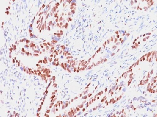
Anti-Rb1 (Tumor Suppressor Protein) Antibody
Mouse Monoclonal Antibody
- 产品详情
- 实验流程
- 背景知识
Application
| WB, IHC-P, IP |
|---|---|
| Primary Accession | P06400 |
| Other Accession | 408528 |
| Reactivity | Human, Mouse |
| Host | Mouse |
| Clonality | Monoclonal |
| Isotype | Mouse / IgG1, kappa |
| Clone Names | SPM353 |
| Calculated MW | 106159 Da |
| Gene ID | 5925 |
|---|---|
| Other Names | OSRC; Osteosarcoma; p105-Rb; PP105; pp110; pRb; Prepro retinoblastoma associated protein; RB1; Retinoblastoma 1; Retinoblastoma-associated protein |
| Application Note | Western Blotting (0.5-1ug/ml); Immunoprecipitation (0.5-1 �g/500ug protein lysate);,Immunohistology (Formalin-fixed) (1-2ug/ml for 30 minutes at RT),(Staining of formalin-fixed tissues requires boiling tissue sections in 10mM citrate buffer, pH 6.0, for 10-20 min followed by cooling at RT for 20 minutes),Optimal dilution for a specific application should be determined. |
| Format | 200ug/ml of Ab purified from Bioreactor Concentrate by Protein A/G. Prepared in 10mM PBS with 0.05% BSA & 0.05% azide. Also available WITHOUT BSA & azide at 1.0mg/ml. |
| Storage | Store at 2 to 8°C.Antibody is stable for 24 months. |
| Precautions | Anti-Rb1 (Tumor Suppressor Protein) Antibody is for research use only and not for use in diagnostic or therapeutic procedures. |
| Name | RB1 |
|---|---|
| Function | Tumor suppressor that is a key regulator of the G1/S transition of the cell cycle (PubMed:10499802). The hypophosphorylated form binds transcription regulators of the E2F family, preventing transcription of E2F-responsive genes (PubMed:10499802). Both physically blocks E2Fs transactivating domain and recruits chromatin- modifying enzymes that actively repress transcription (PubMed:10499802). Cyclin and CDK-dependent phosphorylation of RB1 induces its dissociation from E2Fs, thereby activating transcription of E2F responsive genes and triggering entry into S phase (PubMed:10499802). RB1 also promotes the G0-G1 transition upon phosphorylation and activation by CDK3/cyclin-C (PubMed:15084261). Directly involved in heterochromatin formation by maintaining overall chromatin structure and, in particular, that of constitutive heterochromatin by stabilizing histone methylation. Recruits and targets histone methyltransferases SUV39H1, KMT5B and KMT5C, leading to epigenetic transcriptional repression. Controls histone H4 'Lys-20' trimethylation. Inhibits the intrinsic kinase activity of TAF1. Mediates transcriptional repression by SMARCA4/BRG1 by recruiting a histone deacetylase (HDAC) complex to the c-FOS promoter. In resting neurons, transcription of the c-FOS promoter is inhibited by BRG1- dependent recruitment of a phospho-RB1-HDAC1 repressor complex. Upon calcium influx, RB1 is dephosphorylated by calcineurin, which leads to release of the repressor complex (By similarity). |
| Cellular Location | Nucleus. Cytoplasm {ECO:0000250|UniProtKB:P13405}. Note=During keratinocyte differentiation, acetylation by KAT2B/PCAF is required for nuclear localization (PubMed:20940255). Localizes to the cytoplasm when hyperphosphorylated (By similarity). {ECO:0000250|UniProtKB:P13405, ECO:0000269|PubMed:20940255} |
| Tissue Location | Expressed in the retina. Expressed in foreskin keratinocytes (at protein level) (PubMed:20940255) |
For Research Use Only. Not For Use In Diagnostic Procedures.
Provided below are standard protocols that you may find useful for product applications.
BACKGROUND
Recognizes a 105kDa phosphoprotein, identified as retinoblastoma (Rb) gene product. Its epitope is localized between aa 703-772. It shows no cross reaction with p107 or p130. It specifically stains the nuclei of BT-20 cells and primary human foreskin fibroblast (HFF) cells. It does not stain the Rb-negative BT549 cells. It reacts with the hyperphosphorylated as well as the un (under) phosphorylated form of the Rb protein. Retinoblastoma gene product plays a key role in cell cycle control. It has been identified as a tumor suppressor gene whose loss of its function leads to tumor development. It is widely expressed in a variety of human tissues including breast, esophageal, squamous cell and cervical carcinoma.
终于等到您。ABCEPTA(百远生物)抗体产品。
点击下方“我要评价 ”按钮提交您的反馈信息,您的反馈和评价是我们最宝贵的财富之一,
我们将在1-3个工作日内处理您的反馈信息。
如有疑问,联系:0512-88856768 tech-china@abcepta.com.


