


 癌症的基本特征包括细胞增殖、血管生成、迁移、凋亡逃避机制和细胞永生等。找到癌症发生过程中这些通路的关键标记物和对应的抗体用于检测至关重要。
 为您推荐一个泛素化位点预测神器——泛素化分析工具,可以为您的蛋白的泛素化位点作出预测和评分。
 细胞自噬受体图形绘图工具为你的蛋白的细胞受体结合位点作出预测和评分,识别结合到自噬通路中的蛋白是非常重要的,便于让我们理解自噬在正常生理、病理过程中的作用,如发育、细胞分化、神经退化性疾病、压力条件下、感染和癌症。
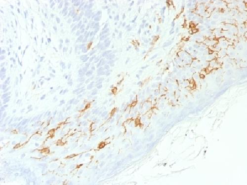
Anti-CD1a / HTA1 Antibody
Recombinant Rabbit Monoclonal Antibody
- 产品详情
- 实验流程
- 背景知识
Application
| IHC-P, IF, FC |
|---|---|
| Primary Accession | P06126 |
| Other Accession | 1309 |
| Reactivity | Human |
| Host | Rabbit |
| Clonality | Monoclonal |
| Isotype | Rabbit / IgG, kappa |
| Clone Names | C1A/1506R |
| Calculated MW | 37077 Da |
| Gene ID | 909 |
|---|---|
| Other Names | Cortical thymocyte antigen CD1A, Epidermal dendritic cell marker CD1a antibody, FCB6, HTA1, T cell surface antigen T6 / Leu 6, T-Cell Surface Glycoprotein CD1A |
| Application Note | Flow Cytometry (0.5-1ug/million cells); Immunofluorescence (1-2ug/ml); ,Immunohistology (Formalin-fixed) (0.5-1ug/ml for 30 min at RT),(Staining of formalin-fixed tissues requires boiling tissue sections in 10mM Citrate Buffer, pH 6.0, for 10-20 min followed by cooling at RT for 20 minutes),Optimal dilution for a specific application should be determined. |
| Format | 200ug/ml of Ab purified by Protein A Column. Prepared in 10mM PBS with 0.05% BSA & 0.05% azide. Also available WITHOUT BSA & azide at 1.0mg/ml. |
| Storage | Store at 2 to 8°C.Antibody is stable for 24 months. |
| Precautions | Anti-CD1a / HTA1 Antibody is for research use only and not for use in diagnostic or therapeutic procedures. |
| Name | CD1A |
|---|---|
| Function | Antigen-presenting protein that binds self and non-self lipid and glycolipid antigens and presents them to T-cell receptors on natural killer T-cells. |
| Cellular Location | Cell membrane; Single-pass type I membrane protein. Membrane raft; Single-pass type I membrane protein. Endosome membrane; Single- pass type I membrane protein. Note=Subject to intracellular trafficking between the cell membrane and endosomes (PubMed:11231314). Localizes to cell surface lipid rafts (PubMed:18178838). |
| Tissue Location | Expressed on cortical thymocytes, epidermal Langerhans cells, dendritic cells, on certain T-cell leukemias, and in various other tissues. |
For Research Use Only. Not For Use In Diagnostic Procedures.
Provided below are standard protocols that you may find useful for product applications.
BACKGROUND
At least five CD1 genes (CD1a, b, c, d, and e) are identified. CD1 proteins have been demonstrated to restrict T cell response to non-peptide lipid and glycolipid antigens and play a role in non-classical antigen presentation. CD1a is a non-polymorphic MHC Class 1 related cell surface glycoprotein, expressed in association with Beta-2 microglobulin. Anti-CD1a labels Langerhans cell histiocytosis (Histiocytosis X), extranodal histiocytic sarcoma, a subset of T-lymphoblastic lymphoma/leukemia, and interdigitating dendritic cell sarcoma of the lymph node. When combined with antibodies against TTF-1 and CD5, anti-CD1a is useful in distinguishing between pulmonary and thymic neoplasms since CD1a is consistently expressed in thymic lymphocytes in both typical and atypical thymomas, but only focally in 1/6 of thymic carcinomas and not in lymphocytes in pulmonary neoplasms. Anti-CD1a is reported to be a new marker for perivascular epithelial cell tumor (PEComa).
终于等到您。ABCEPTA(百远生物)抗体产品。
点击下方“我要评价 ”按钮提交您的反馈信息,您的反馈和评价是我们最宝贵的财富之一,
我们将在1-3个工作日内处理您的反馈信息。
如有疑问,联系:0512-88856768 tech-china@abcepta.com.


