


 癌症的基本特征包括细胞增殖、血管生成、迁移、凋亡逃避机制和细胞永生等。找到癌症发生过程中这些通路的关键标记物和对应的抗体用于检测至关重要。
 为您推荐一个泛素化位点预测神器——泛素化分析工具,可以为您的蛋白的泛素化位点作出预测和评分。
 细胞自噬受体图形绘图工具为你的蛋白的细胞受体结合位点作出预测和评分,识别结合到自噬通路中的蛋白是非常重要的,便于让我们理解自噬在正常生理、病理过程中的作用,如发育、细胞分化、神经退化性疾病、压力条件下、感染和癌症。
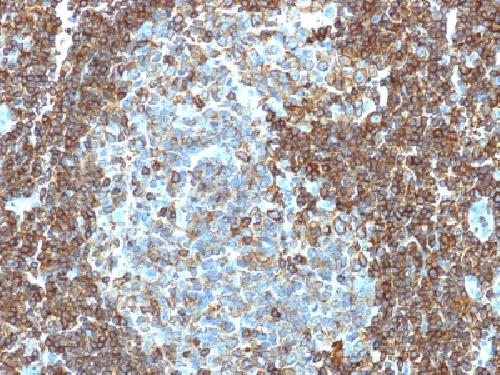
Anti-CD3e (T-Cell Marker) Antibody
Mouse Monoclonal Antibody
- 产品详情
- 实验流程
- 背景知识
Application
| WB, IHC-P, IF, FC |
|---|---|
| Primary Accession | P07766 |
| Other Accession | 3003 |
| Reactivity | Human |
| Host | Mouse |
| Clonality | Monoclonal |
| Isotype | Mouse / IgG1 |
| Clone Names | PC3/188A |
| Calculated MW | 23147 Da |
| Gene ID | 916 |
|---|---|
| Other Names | CD 3E, CD3 epsilon, CD3 TCR complex, CD3E, CD3e antigen epsilon polypeptide (TiT3 complex), T cell antigen receptor complex epsilon subunit of T3, T-cell surface antigen T3/Leu-4 epsilon chain, T-cell surface glycoprotein CD3 epsilon chain, T3E, TCRE, TiT3 complex |
| Application Note | Flow Cytometry (0.5-1ug/million cells); ,Immunofluorescence (1-2ug/ml); ,Western Blotting (1-2ug/ml for 60 minutes at RT);,Immunohistology (Formalin-fixed) (1-2ug/ml for 30 minutes at RT),(Staining of formalin-fixed tissues requires boiling tissue sections in 10mM citrate buffer, pH 6.0, for 10-20 min followed by cooling at RT for 20 minutes),Optimal dilution for a specific application should be determined. |
| Format | 200ug/ml of Ab purified from rabbit anti-serum by Protein A. Prepared in 10mM PBS with 0.05% BSA & 0.05% azide. Also available WITHOUT BSA at 1.0mg/ml. |
| Storage | Store at 2 to 8°C.Antibody is stable for 24 months. |
| Precautions | Anti-CD3e (T-Cell Marker) Antibody is for research use only and not for use in diagnostic or therapeutic procedures. |
| Name | CD3E |
|---|---|
| Synonyms | T3E |
| Function | Part of the TCR-CD3 complex present on T-lymphocyte cell surface that plays an essential role in adaptive immune response. When antigen presenting cells (APCs) activate T-cell receptor (TCR), TCR- mediated signals are transmitted across the cell membrane by the CD3 chains CD3D, CD3E, CD3G and CD3Z. All CD3 chains contain immunoreceptor tyrosine-based activation motifs (ITAMs) in their cytoplasmic domain. Upon TCR engagement, these motifs become phosphorylated by Src family protein tyrosine kinases LCK and FYN, resulting in the activation of downstream signaling pathways (PubMed:2470098). In addition of this role of signal transduction in T-cell activation, CD3E plays an essential role in correct T-cell development. Initiates the TCR-CD3 complex assembly by forming the two heterodimers CD3D/CD3E and CD3G/CD3E. Also participates in internalization and cell surface down- regulation of TCR-CD3 complexes via endocytosis sequences present in CD3E cytosolic region (PubMed:10384095, PubMed:26507128). In addition to its role as a TCR coreceptor, it serves as a receptor for ITPRIPL1. Ligand recognition inhibits T-cell activation by promoting interaction with NCK1, which prevents CD3E-ZAP70 interaction and blocks the ERK- NFkB signaling cascade and calcium influx (PubMed:38614099). |
| Cellular Location | Cell membrane; Single-pass type I membrane protein |
For Research Use Only. Not For Use In Diagnostic Procedures.
Provided below are standard protocols that you may find useful for product applications.
BACKGROUND
Recognizes the -chain of CD3, which consists of five different polypeptide chains (designated as gamma, delta, epsilon, zeta, and eta) with MW ranging from 16-28kDa. The CD3 complex is closely associated at the lymphocyte cell surface with the T cell antigen receptor (TCR). Reportedly, CD3 complex is involved in signal transduction to the T cell interior following antigen recognition. The CD3 antigen is first detectable in early thymocytes and probably represents one of the earliest signs of commitment to the T cell lineage. In cortical thymocytes, CD3 is predominantly intra-cytoplasmic. However, in medullary thymocytes, it appears on the T cell surface. CD3 antigen is a highly specific marker for T cells, and is present in majority of T cell neoplasms.
终于等到您。ABCEPTA(百远生物)抗体产品。
点击下方“我要评价 ”按钮提交您的反馈信息,您的反馈和评价是我们最宝贵的财富之一,
我们将在1-3个工作日内处理您的反馈信息。
如有疑问,联系:0512-88856768 tech-china@abcepta.com.


