


 癌症的基本特征包括细胞增殖、血管生成、迁移、凋亡逃避机制和细胞永生等。找到癌症发生过程中这些通路的关键标记物和对应的抗体用于检测至关重要。
 为您推荐一个泛素化位点预测神器——泛素化分析工具,可以为您的蛋白的泛素化位点作出预测和评分。
 细胞自噬受体图形绘图工具为你的蛋白的细胞受体结合位点作出预测和评分,识别结合到自噬通路中的蛋白是非常重要的,便于让我们理解自噬在正常生理、病理过程中的作用,如发育、细胞分化、神经退化性疾病、压力条件下、感染和癌症。
DYRK3 antibody - N-terminal region
Rabbit Polyclonal Antibody
- 产品详情
- 实验流程
- 背景知识
Application
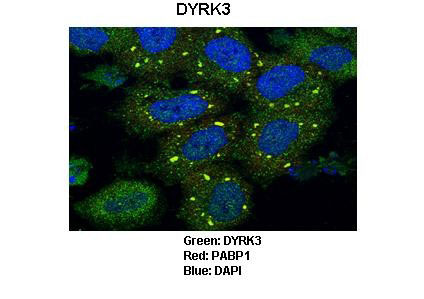
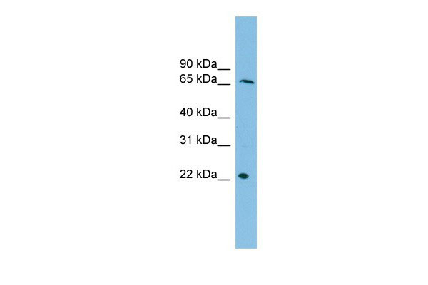
| WB, IF |
|---|---|
| Primary Accession | O43781 |
| Other Accession | O43781, NP_003573, NM_003582 |
| Reactivity | Human, Mouse |
| Predicted | Human, Mouse |
| Host | Rabbit |
| Clonality | Polyclonal |
| Calculated MW | 65714 Da |
| Gene ID | 8444 |
|---|---|
| Alias Symbol | DYRK5, RED, REDK, hYAK3-2 |
| Other Names | Dual specificity tyrosine-phosphorylation-regulated kinase 3, Regulatory erythroid kinase, REDK, DYRK3 |
| Target/Specificity | This gene product belongs to the DYRK family of dual-specificity protein kinases that catalyze autophosphorylation on serine/threonine and tyrosine residues. The members of this family share structural similarity, however, differ in their substrate specificity, suggesting their involvement in different cellular functions. The encoded protein has been shown to autophosphorylate on tyrosine residue and catalyze phosphorylation of histones H3 and H2B in vitro. Alternatively spliced transcript variants encoding different isoforms have been identified. |
| Format | Liquid. Purified antibody supplied in 1x PBS buffer with 0.09% (w/v) sodium azide and 2% sucrose. |
| Reconstitution & Storage | Add 50 ul of distilled water. Final anti-DYRK3 antibody concentration is 1 mg/ml in PBS buffer with 2% sucrose. For longer periods of storage, store at -20°C. Avoid repeat freeze-thaw cycles. |
| Precautions | DYRK3 antibody - N-terminal region is for research use only and not for use in diagnostic or therapeutic procedures. |
| Name | DYRK3 (HGNC:3094) |
|---|---|
| Function | Dual-specificity protein kinase that promotes disassembly of several types of membraneless organelles during mitosis, such as stress granules, nuclear speckles and pericentriolar material (PubMed:29973724). Dual-specificity tyrosine-regulated kinases (DYRKs) autophosphorylate a critical tyrosine residue in their activation loop and phosphorylate their substrate on serine and threonine residues (PubMed:29634919, PubMed:9748265). Acts as a central dissolvase of membraneless organelles during the G2-to-M transition, after the nuclear-envelope breakdown: acts by mediating phosphorylation of multiple serine and threonine residues in unstructured domains of proteins, such as SRRM1 and PCM1 (PubMed:29973724). Does not mediate disassembly of all membraneless organelles: disassembly of P-body and nucleolus is not regulated by DYRK3 (PubMed:29973724). Dissolution of membraneless organelles at the onset of mitosis is also required to release mitotic regulators, such as ZNF207, from liquid-unmixed organelles where they are sequestered and keep them dissolved during mitosis (PubMed:29973724). Regulates mTORC1 by mediating the dissolution of stress granules: during stressful conditions, DYRK3 partitions from the cytosol to the stress granule, together with mTORC1 components, which prevents mTORC1 signaling (PubMed:23415227). When stress signals are gone, the kinase activity of DYRK3 is required for the dissolution of stress granule and mTORC1 relocation to the cytosol: acts by mediating the phosphorylation of the mTORC1 inhibitor AKT1S1, allowing full reactivation of mTORC1 signaling (PubMed:23415227). Also acts as a negative regulator of EPO-dependent erythropoiesis: may place an upper limit on red cell production during stress erythropoiesis (PubMed:10779429). Inhibits cell death due to cytokine withdrawal in hematopoietic progenitor cells (PubMed:10779429). Promotes cell survival upon genotoxic stress through phosphorylation of SIRT1: this in turn inhibits p53/TP53 activity and apoptosis (PubMed:20167603). |
| Cellular Location | Nucleus. Cytoplasm. Nucleus speckle. Cytoplasmic granule. Cytoplasm, cytoskeleton, microtubule organizing center, centrosome Note=Associates with membraneless organelles in the cytoplasm and nucleus (PubMed:29973724). Shuttles between cytoplasm and stress granules (PubMed:20167603). Localized predominantly on distinct speckles distributed throughout the cytoplasm of the cell (PubMed:20167603). At low concentration, showns a homogeneous distribution throughout the cytoplasm and does not condense in speckles. During oxidative and osmotic stress, localizes to stress granules (PubMed:20167603). |
| Tissue Location | Isoform 1: Highly expressed in testis and in hematopoietic tissue such as fetal liver, and bone marrow (PubMed:10779429). Isoform 1: Predominant form in fetal liver and bone marrow (PubMed:10779429). Isoform 1: Present at low levels in heart, pancreas, lymph node and thymus (PubMed:10779429). Isoform 2: Highly expressed in testis and in hematopoietic tissue such as fetal liver, and bone marrow (PubMed:10779429). Isoform 2: Predominant form in testis. Isoform 2: Present at low levels in heart, pancreas, lymph node and thymus (PubMed:10779429). |
For Research Use Only. Not For Use In Diagnostic Procedures.
Provided below are standard protocols that you may find useful for product applications.
BACKGROUND
This is a rabbit polyclonal antibody against DYRK3. It was validated on Western Blot using a cell lysate as a positive control. Abgent strives to provide antibodies covering each member of a whole protein family of your interest. We also use our best efforts to provide you antibodies recognize various epitopes of a target protein. For availability of antibody needed for your experiment, please inquire (sales@abgent.com).
终于等到您。ABCEPTA(百远生物)抗体产品。
点击下方“我要评价 ”按钮提交您的反馈信息,您的反馈和评价是我们最宝贵的财富之一,
我们将在1-3个工作日内处理您的反馈信息。
如有疑问,联系:0512-88856768 tech-china@abcepta.com.


