


 癌症的基本特征包括细胞增殖、血管生成、迁移、凋亡逃避机制和细胞永生等。找到癌症发生过程中这些通路的关键标记物和对应的抗体用于检测至关重要。
 为您推荐一个泛素化位点预测神器——泛素化分析工具,可以为您的蛋白的泛素化位点作出预测和评分。
 细胞自噬受体图形绘图工具为你的蛋白的细胞受体结合位点作出预测和评分,识别结合到自噬通路中的蛋白是非常重要的,便于让我们理解自噬在正常生理、病理过程中的作用,如发育、细胞分化、神经退化性疾病、压力条件下、感染和癌症。
SIAH1 antibody - N-terminal region
Rabbit Polyclonal Antibody
- 产品详情
- 实验流程
- 背景知识
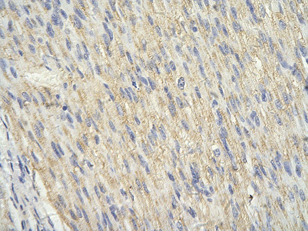
Application
| WB, IHC |
|---|---|
| Primary Accession | Q8IUQ4 |
| Other Accession | Q8IUQ4-2, NP_001006611, NM_001006610 |
| Reactivity | Human, Mouse, Rat, Rabbit, Zebrafish, Dog, Guinea Pig, Horse, Bovine |
| Predicted | Human, Mouse, Rat, Zebrafish, Chicken, Dog, Guinea Pig, Bovine |
| Host | Rabbit |
| Clonality | Polyclonal |
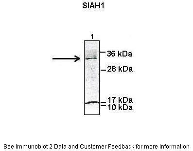
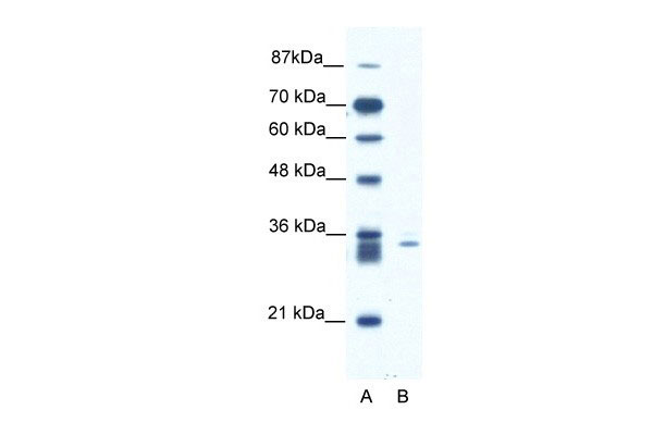
| Calculated MW | 31123 Da |
| Gene ID | 6477 |
|---|---|
| Alias Symbol | SIAH1A |
| Other Names | E3 ubiquitin-protein ligase SIAH1, 632-, Seven in absentia homolog 1, Siah-1, Siah-1a, SIAH1, HUMSIAH |
| Target/Specificity | SIAH1 is a protein that is a member of the seven in absentia homolog (SIAH) family. The protein is an E3 ligase and is involved in ubiquitination and proteasome-mediated degradation of specific proteins. The activity of this ubiquitin ligase has been implicated in the development of certain forms of Parkinson's disease, the regulation of the cellular response to hypoxia and induction of apoptosis. Alternative splicing results in several additional transcript variants, some encoding different isoforms and others that have not been fully characterized. |
| Format | Liquid. Purified antibody supplied in 1x PBS buffer with 0.09% (w/v) sodium azide and 2% sucrose. |
| Reconstitution & Storage | Add 100 ul of distilled water. Final anti-SIAH1 antibody concentration is 1 mg/ml in PBS buffer with 2% sucrose. For longer periods of storage, store at -20°C. Avoid repeat freeze-thaw cycles. |
| Precautions | SIAH1 antibody - N-terminal region is for research use only and not for use in diagnostic or therapeutic procedures. |
| Name | SIAH1 |
|---|---|
| Synonyms | HUMSIAH |
| Function | E3 ubiquitin-protein ligase that mediates ubiquitination and subsequent proteasomal degradation of target proteins (PubMed:14506261, PubMed:14645235, PubMed:14654780, PubMed:15064394, PubMed:16085652, PubMed:19224863, PubMed:20508617, PubMed:22483617, PubMed:28546513, PubMed:32430360, PubMed:33591310, PubMed:9334332, PubMed:9858595). E3 ubiquitin ligases accept ubiquitin from an E2 ubiquitin-conjugating enzyme in the form of a thioester and then directly transfers the ubiquitin to targeted substrates (PubMed:14506261, PubMed:14645235, PubMed:14654780, PubMed:15064394, PubMed:16085652, PubMed:19224863, PubMed:20508617, PubMed:22483617, PubMed:9334332, PubMed:9858595). Mediates E3 ubiquitin ligase activity either through direct binding to substrates or by functioning as the essential RING domain subunit of larger E3 complexes (PubMed:14506261, PubMed:14645235, PubMed:14654780, PubMed:15064394, PubMed:16085652, PubMed:19224863, PubMed:20508617, PubMed:22483617, PubMed:9334332, PubMed:9858595). Triggers the ubiquitin-mediated degradation of many substrates, including proteins involved in transcription regulation (ELL2, MYB, POU2AF1, PML and RBBP8), a cell surface receptor (DCC), the cell-surface receptor-type tyrosine kinase FLT3, the cytoplasmic signal transduction molecules (KLF10/TIEG1 and NUMB), an antiapoptotic protein (BAG1), a microtubule motor protein (KIF22), a protein involved in synaptic vesicle function in neurons (SYP), a structural protein (CTNNB1) and SNCAIP (PubMed:10747903, PubMed:11146551, PubMed:11389839, PubMed:11389840, PubMed:11483517, PubMed:11483518, PubMed:11752454, PubMed:12072443). Confers constitutive instability to HIPK2 through proteasomal degradation (PubMed:18536714, PubMed:33591310). It is thereby involved in many cellular processes such as apoptosis, tumor suppression, cell cycle, axon guidance, transcription regulation, spermatogenesis and TNF-alpha signaling (PubMed:14506261, PubMed:14645235, PubMed:14654780, PubMed:15064394, PubMed:16085652, PubMed:19224863, PubMed:20508617, PubMed:22483617, PubMed:9334332, PubMed:9858595). Has some overlapping function with SIAH2 (PubMed:14506261, PubMed:14645235, PubMed:14654780, PubMed:15064394, PubMed:16085652, PubMed:19224863, PubMed:20508617, PubMed:22483617, PubMed:9334332, PubMed:9858595). Induces apoptosis in cooperation with PEG3 (By similarity). Upon nitric oxid (NO) generation that follows apoptotic stimulation, interacts with S-nitrosylated GAPDH, mediating the translocation of GAPDH to the nucleus (By similarity). GAPDH acts as a stabilizer of SIAH1, facilitating the degradation of nuclear proteins (By similarity). Mediates ubiquitination and degradation of EGLN2 and EGLN3 in response to the unfolded protein response (UPR), leading to their degradation and subsequent stabilization of ATF4 (By similarity). Also part of the Wnt signaling pathway in which it mediates the Wnt-induced ubiquitin- mediated proteasomal degradation of AXIN1 (PubMed:28546513, PubMed:32430360). |
| Cellular Location | Cytoplasm. Nucleus. Note=Predominantly cytoplasmic. Partially nuclear |
| Tissue Location | Widely expressed at a low level. Down-regulated in advanced hepatocellular carcinomas. |
For Research Use Only. Not For Use In Diagnostic Procedures.
Provided below are standard protocols that you may find useful for product applications.
BACKGROUND
This is a rabbit polyclonal antibody against SIAH1. It was validated on Western Blot using a cell lysate as a positive control. Abgent strives to provide antibodies covering each member of a whole protein family of your interest. We also use our best efforts to provide you antibodies recognize various epitopes of a target protein. For availability of antibody needed for your experiment, please inquire (sales@abgent.com).
终于等到您。ABCEPTA(百远生物)抗体产品。
点击下方“我要评价 ”按钮提交您的反馈信息,您的反馈和评价是我们最宝贵的财富之一,
我们将在1-3个工作日内处理您的反馈信息。
如有疑问,联系:0512-88856768 tech-china@abcepta.com.


