


 癌症的基本特征包括细胞增殖、血管生成、迁移、凋亡逃避机制和细胞永生等。找到癌症发生过程中这些通路的关键标记物和对应的抗体用于检测至关重要。
 为您推荐一个泛素化位点预测神器——泛素化分析工具,可以为您的蛋白的泛素化位点作出预测和评分。
 细胞自噬受体图形绘图工具为你的蛋白的细胞受体结合位点作出预测和评分,识别结合到自噬通路中的蛋白是非常重要的,便于让我们理解自噬在正常生理、病理过程中的作用,如发育、细胞分化、神经退化性疾病、压力条件下、感染和癌症。
BRD2 antibody - C-terminal region
Rabbit Polyclonal Antibody
- 产品详情
- 实验流程
- 背景知识
Application
| WB |
|---|---|
| Primary Accession | P25440 |
| Other Accession | P25440, NP_005095, NM_005104 |
| Reactivity | Human, Mouse, Rat, Dog, Guinea Pig, Horse, Bovine |
| Predicted | Human, Mouse, Rat, Pig, Chicken, Dog, Guinea Pig, Bovine |
| Host | Rabbit |
| Clonality | Polyclonal |
| Calculated MW | 88061 Da |
| Gene ID | 6046 |
|---|---|
| Alias Symbol | FSH, NAT, RNF3, FSRG1, RING3, D6S113E |
| Other Names | Bromodomain-containing protein 2, O2711, Really interesting new gene 3 protein, BRD2, KIAA9001, RING3 |
| Target/Specificity | BRD2 is a mitogen-activated kinase which localizes to the nucleus. The gene maps to the major histocompatability complex (MHC) class II region on chromosome 6p21.3 but sequence comparison suggests that the protein is not involved in the immune response. Homology to the Drosophila gene female sterile homeotic suggests that this human gene may be part of a signal transduction pathway involved in growth control. |
| Format | Liquid. Purified antibody supplied in 1x PBS buffer with 0.09% (w/v) sodium azide and 2% sucrose. |
| Reconstitution & Storage | Add 100 ul of distilled water. Final anti-BRD2 antibody concentration is 1 mg/ml in PBS buffer with 2% sucrose. For longer periods of storage, store at -20°C. Avoid repeat freeze-thaw cycles. |
| Precautions | BRD2 antibody - C-terminal region is for research use only and not for use in diagnostic or therapeutic procedures. |
| Name | BRD2 {ECO:0000303|PubMed:16227282, ECO:0000312|HGNC:HGNC:1103} |
|---|---|
| Function | Chromatin reader protein that specifically recognizes and binds histone H4 acetylated at 'Lys-5' and 'Lys-12' (H4K5ac and H4K12ac, respectively), thereby controlling gene expression and remodeling chromatin structures (PubMed:17148447, PubMed:17848202, PubMed:18406326, PubMed:20048151, PubMed:20709061, PubMed:20871596). Recruits transcription factors and coactivators to target gene sites, and activates RNA polymerase II machinery for transcriptional elongation (PubMed:28262505). Plays a key role in genome compartmentalization via its association with CTCF and cohesin: recruited to chromatin by CTCF and promotes formation of topologically associating domains (TADs) via its ability to bind acetylated histones, contributing to CTCF boundary formation and enhancer insulation (PubMed:35410381). Also recognizes and binds acetylated non-histone proteins, such as STAT3 (PubMed:28262505). Involved in inflammatory response by regulating differentiation of naive CD4(+) T-cells into T- helper Th17: recognizes and binds STAT3 acetylated at 'Lys-87', promoting STAT3 recruitment to chromatin (PubMed:28262505). In addition to acetylated lysines, also recognizes and binds lysine residues on histones that are both methylated and acetylated on the same side chain to form N6-acetyl-N6-methyllysine (Kacme), an epigenetic mark of active chromatin associated with increased transcriptional initiation (PubMed:37731000). Specifically binds histone H4 acetyl-methylated at 'Lys-5' and 'Lys-12' (H4K5acme and H4K12acme, respectively) (PubMed:37731000). |
| Cellular Location | Nucleus. Chromosome Note=Detected on chromatin and nucleosomes |
For Research Use Only. Not For Use In Diagnostic Procedures.
Provided below are standard protocols that you may find useful for product applications.
BACKGROUND
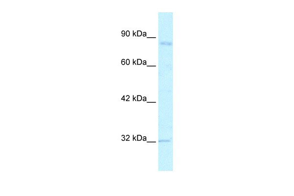
This is a rabbit polyclonal antibody against BRD2. It was validated on Western Blot using a cell lysate as a positive control. Abgent strives to provide antibodies covering each member of a whole protein family of your interest. We also use our best efforts to provide you antibodies recognize various epitopes of a target protein. For availability of antibody needed for your experiment, please inquire (sales@abgent.com).
终于等到您。ABCEPTA(百远生物)抗体产品。
点击下方“我要评价 ”按钮提交您的反馈信息,您的反馈和评价是我们最宝贵的财富之一,
我们将在1-3个工作日内处理您的反馈信息。
如有疑问,联系:0512-88856768 tech-china@abcepta.com.


