


 癌症的基本特征包括细胞增殖、血管生成、迁移、凋亡逃避机制和细胞永生等。找到癌症发生过程中这些通路的关键标记物和对应的抗体用于检测至关重要。
 为您推荐一个泛素化位点预测神器——泛素化分析工具,可以为您的蛋白的泛素化位点作出预测和评分。
 细胞自噬受体图形绘图工具为你的蛋白的细胞受体结合位点作出预测和评分,识别结合到自噬通路中的蛋白是非常重要的,便于让我们理解自噬在正常生理、病理过程中的作用,如发育、细胞分化、神经退化性疾病、压力条件下、感染和癌症。
RIPX antibody - C-terminal region
Rabbit Polyclonal Antibody
- 产品详情
- 实验流程
- 背景知识
Application
| WB |
|---|---|
| Primary Accession | Q7L099 |
| Other Accession | Q7L099, NP_055776, NM_014961 |
| Reactivity | Human, Mouse, Rat, Rabbit, Pig, Dog, Guinea Pig, Horse, Bovine |
| Predicted | Human, Mouse, Rat, Dog, Bovine |
| Host | Rabbit |
| Clonality | Polyclonal |
| Calculated MW | 52965 Da |
| Gene ID | 22902 |
|---|---|
| Alias Symbol | RIPX, SINGAR1 |
| Other Names | Protein RUFY3, Rap2-interacting protein x, RIPx, Single axon-regulated protein, Singar, RUFY3, KIAA0871 |
| Target/Specificity | Located on chromosome 4, the RIPX encodes a protein with unknown function. |
| Format | Liquid. Purified antibody supplied in 1x PBS buffer with 0.09% (w/v) sodium azide and 2% sucrose. |
| Reconstitution & Storage | Add 50 ul of distilled water. Final anti-RIPX antibody concentration is 1 mg/ml in PBS buffer with 2% sucrose. For longer periods of storage, store at -20°C. Avoid repeat freeze-thaw cycles. |
| Precautions | RIPX antibody - C-terminal region is for research use only and not for use in diagnostic or therapeutic procedures. |
| Name | RUFY3 (HGNC:30285) |
|---|---|
| Synonyms | KIAA0871 |
| Function | ARL8 effector that promotes the coupling of endolysosomes to dynein-dynactin for retrograde transport along microtubules. Acts by binding both GTP-bound ARL8 and dynein-dynactin. In nonneuronal cells, promotes concentration of endolysosomes in the juxtanuclear area. In hippocampal neurons, drives retrograde transport of endolysosomes from the axon to the soma (PubMed:35314674). Plays a role in the generation of neuronal polarity formation and axon growth (By similarity). Implicated in the formation of a single axon by developing neurons (By similarity). May inhibit the formation of additional axons by inhibition of PI3K in minor neuronal processes (By similarity). Plays a role in the formation of F-actin-enriched protrusive structures at the cell periphery (PubMed:25766321). Plays a role in cytoskeletal organization by regulating the subcellular localization of FSCN1 and DBN1 at axonal growth cones (By similarity). |
| Cellular Location | Cytoplasm. Endomembrane system. Cell projection, invadopodium. Perikaryon {ECO:0000250|UniProtKB:Q9D394}. Cell projection {ECO:0000250|UniProtKB:Q9D394}. Cell projection, growth cone {ECO:0000250|UniProtKB:Q9D394}. Cell projection, filopodium {ECO:0000250|UniProtKB:Q9D394}. Cell projection, lamellipodium {ECO:0000250|UniProtKB:Q9D394}. Lysosome Note=Colocalizes with PAK1, F-actin, myosins and integrins in invadopodia at the cell periphery (PubMed:25766321). Colocalizes with Ras-related Rab-5 proteins in cytoplasmic vesicles (PubMed:20376209) Accumulates in axon growth cones in a F-actin-dependent manner (By similarity). Colocalizes with FSCN1 and F-actin at filipodia and lamellipodia of axonal growth cones (By similarity). Colocalizes with DBN1 and F-actin at transitional domain of the axonal growth cone (By similarity). Recruitment to endolysosomes partially depends upon the presence of ARL8 (PubMed:35314674). {ECO:0000250|UniProtKB:Q5FVJ0, ECO:0000250|UniProtKB:Q9D394, ECO:0000269|PubMed:20376209, ECO:0000269|PubMed:25766321, ECO:0000269|PubMed:35314674} |
| Tissue Location | Overexpressed in gastric cancer cells and tissues (at protein level) (PubMed:25766321). |
For Research Use Only. Not For Use In Diagnostic Procedures.
Provided below are standard protocols that you may find useful for product applications.
BACKGROUND
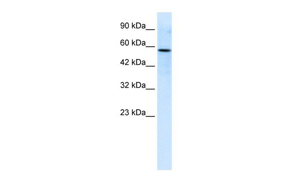
This is a rabbit polyclonal antibody against RIPX. It was validated on Western Blot using a cell lysate as a positive control. Abgent strives to provide antibodies covering each member of a whole protein family of your interest. We also use our best efforts to provide you antibodies recognize various epitopes of a target protein. For availability of antibody needed for your experiment, please inquire (sales@abgent.com).
终于等到您。ABCEPTA(百远生物)抗体产品。
点击下方“我要评价 ”按钮提交您的反馈信息,您的反馈和评价是我们最宝贵的财富之一,
我们将在1-3个工作日内处理您的反馈信息。
如有疑问,联系:0512-88856768 tech-china@abcepta.com.


