


 癌症的基本特征包括细胞增殖、血管生成、迁移、凋亡逃避机制和细胞永生等。找到癌症发生过程中这些通路的关键标记物和对应的抗体用于检测至关重要。
 为您推荐一个泛素化位点预测神器——泛素化分析工具,可以为您的蛋白的泛素化位点作出预测和评分。
 细胞自噬受体图形绘图工具为你的蛋白的细胞受体结合位点作出预测和评分,识别结合到自噬通路中的蛋白是非常重要的,便于让我们理解自噬在正常生理、病理过程中的作用,如发育、细胞分化、神经退化性疾病、压力条件下、感染和癌症。
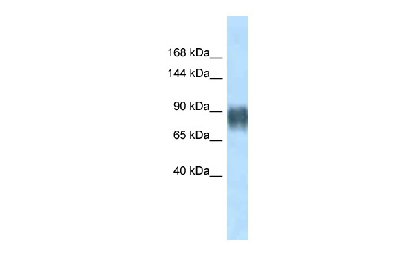
Avil antibody - N-terminal region
Rabbit Polyclonal Antibody
- 产品详情
- 实验流程
Application
| WB |
|---|---|
| Primary Accession | Q9WU06 |
| Other Accession | NM_024401, NP_077377 |
| Reactivity | Human, Mouse, Rat, Zebrafish, Dog, Horse, Bovine |
| Predicted | Human, Mouse, Rat, Dog, Guinea Pig, Bovine |
| Host | Rabbit |
| Clonality | Polyclonal |
| Calculated MW | 91894 Da |
| Gene ID | 79253 |
|---|---|
| Other Names | Advillin, Peripheral nervous system villin-like protein, Pervin, Avil, Advil |
| Format | Liquid. Purified antibody supplied in 1x PBS buffer with 0.09% (w/v) sodium azide and 2% sucrose. |
| Reconstitution & Storage | Add 50 ul of distilled water. Final anti-Avil antibody concentration is 1 mg/ml in PBS buffer with 2% sucrose. For longer periods of storage, store at 20°C. Avoid repeat freeze-thaw cycles. |
| Precautions | Avil antibody - N-terminal region is for research use only and not for use in diagnostic or therapeutic procedures. |
| Name | Avil {ECO:0000312|RGD:620301} |
|---|---|
| Function | Ca(2+)-regulated actin-binding protein which plays an important role in actin bundling. May have a unique function in the morphogenesis of neuronal cells which form ganglia. Required for SREC1- mediated regulation of neurite-like outgrowth. Plays a role in regenerative sensory axon outgrowth and remodeling processes after peripheral injury in neonates (PubMed:11849295). Involved in the formation of long fine actin-containing filopodia-like structures in fibroblast. Plays a role in ciliogenesis. In podocytes, controls lamellipodia formation through the regulation of EGF-induced diacylglycerol generation by PLCE1 and ARP2/3 complex assembly (By similarity). |
| Cellular Location | Cytoplasm, cytoskeleton. Cell projection, neuron projection. Cell projection, axon. Cell projection, lamellipodium {ECO:0000250|UniProtKB:O75366}. Cell junction, focal adhesion {ECO:0000250|UniProtKB:O75366}. Note=In podocytes, present in the F- actin-enriched cell periphery that generates lamellipodia and focal adhesions. {ECO:0000250|UniProtKB:O75366} |
| Tissue Location | Expressed in dorsal root ganglion (DRG) neurons and superior cervical ganglia (SCG) (PubMed:11849295). Expressed in podocytes (PubMed:29058690). |
Research Areas
For Research Use Only. Not For Use In Diagnostic Procedures.
Application Protocols
Provided below are standard protocols that you may find useful for product applications.
终于等到您。ABCEPTA(百远生物)抗体产品。
点击下方“我要评价 ”按钮提交您的反馈信息,您的反馈和评价是我们最宝贵的财富之一,
我们将在1-3个工作日内处理您的反馈信息。
如有疑问,联系:0512-88856768 tech-china@abcepta.com.


