


 癌症的基本特征包括细胞增殖、血管生成、迁移、凋亡逃避机制和细胞永生等。找到癌症发生过程中这些通路的关键标记物和对应的抗体用于检测至关重要。
 为您推荐一个泛素化位点预测神器——泛素化分析工具,可以为您的蛋白的泛素化位点作出预测和评分。
 细胞自噬受体图形绘图工具为你的蛋白的细胞受体结合位点作出预测和评分,识别结合到自噬通路中的蛋白是非常重要的,便于让我们理解自噬在正常生理、病理过程中的作用,如发育、细胞分化、神经退化性疾病、压力条件下、感染和癌症。
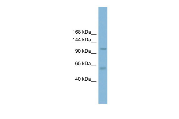
CLCA2 antibody - C-terminal region
Rabbit Polyclonal Antibody
- 产品详情
- 实验流程
Application
| WB |
|---|---|
| Primary Accession | Q9UQC9 |
| Other Accession | NM_006536, NP_006527 |
| Reactivity | Human, Mouse, Rat, Rabbit, Pig, Dog, Horse, Bovine |
| Predicted | Human, Rabbit, Pig, Dog, Horse, Bovine |
| Host | Rabbit |
| Clonality | Polyclonal |
| Calculated MW | 103941 Da |
| Gene ID | 9635 |
|---|---|
| Alias Symbol | CACC, CACC3, CLCRG2, CaCC-3, FLJ97885 |
| Other Names | Calcium-activated chloride channel regulator 2, 3.4.-.-, Calcium-activated chloride channel family member 2, hCLCA2, Calcium-activated chloride channel protein 3, CaCC-3, hCaCC-3, Calcium-activated chloride channel regulator 2, 109 kDa form, Calcium-activated chloride channel regulator 2, 35 kDa form, CLCA2, CACC3 |
| Format | Liquid. Purified antibody supplied in 1x PBS buffer with 0.09% (w/v) sodium azide and 2% sucrose. |
| Reconstitution & Storage | Add 50 ul of distilled water. Final anti-CLCA2 antibody concentration is 1 mg/ml in PBS buffer with 2% sucrose. For longer periods of storage, store at 20°C. Avoid repeat freeze-thaw cycles. |
| Precautions | CLCA2 antibody - C-terminal region is for research use only and not for use in diagnostic or therapeutic procedures. |
| Name | CLCA2 |
|---|---|
| Synonyms | CACC3 |
| Function | Plays a role in modulating chloride current across the plasma membrane in a calcium-dependent manner, and cell adhesion. Involved in basal cell adhesion and/or stratification of squamous epithelia. May act as a tumor suppressor in breast and colorectal cancer. Plays a key role for cell adhesion in the beginning stages of lung metastasis via the binding to ITGB4. |
| Cellular Location | Cell membrane; Single-pass type I membrane protein. Basal cell membrane; Single-pass type I membrane protein. Cell junction |
| Tissue Location | Expressed in cornea, skin, vagina, esophagus, and larynx (at protein level). Expressed in trachea and mammary gland Weakly expressed in testis and kidney. Highly expressed in corneal epithelium, colon and trachea. Moderately expressed in brain, urogenital organs, bladder, uterus and prostate. Highly expressed in tissues containing stratified epithelium including cornea, esophagus, larynx, skin and vagina than those tissues which contain only epithelial monolayers. Expressed in normal breast epithelium but not in breast cancer. Highly expressed during epithelial stratification Expressed in endothelial cells of lung. Expressed selectively in endothelia of small pulmonary arteries, arterioles, and subpleural and interlobular venules. |
Research Areas
For Research Use Only. Not For Use In Diagnostic Procedures.
Application Protocols
Provided below are standard protocols that you may find useful for product applications.
终于等到您。ABCEPTA(百远生物)抗体产品。
点击下方“我要评价 ”按钮提交您的反馈信息,您的反馈和评价是我们最宝贵的财富之一,
我们将在1-3个工作日内处理您的反馈信息。
如有疑问,联系:0512-88856768 tech-china@abcepta.com.


