


 癌症的基本特征包括细胞增殖、血管生成、迁移、凋亡逃避机制和细胞永生等。找到癌症发生过程中这些通路的关键标记物和对应的抗体用于检测至关重要。
 为您推荐一个泛素化位点预测神器——泛素化分析工具,可以为您的蛋白的泛素化位点作出预测和评分。
 细胞自噬受体图形绘图工具为你的蛋白的细胞受体结合位点作出预测和评分,识别结合到自噬通路中的蛋白是非常重要的,便于让我们理解自噬在正常生理、病理过程中的作用,如发育、细胞分化、神经退化性疾病、压力条件下、感染和癌症。
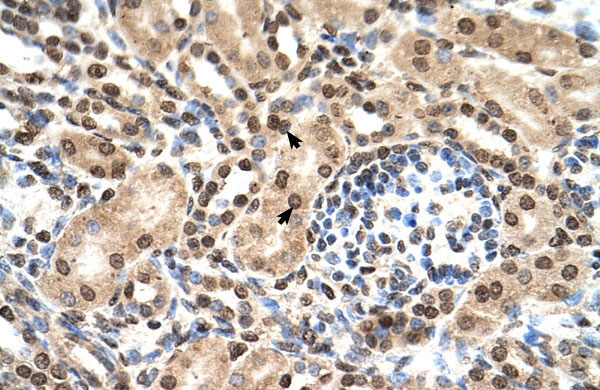
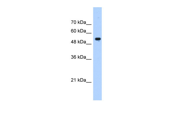
IFIT3 antibody - N-terminal region
Rabbit Polyclonal Antibody
- 产品详情
- 实验流程
Application
| WB, IHC |
|---|---|
| Primary Accession | O14879 |
| Other Accession | NM_001031683, NP_001026853 |
| Reactivity | Human, Mouse, Rat, Rabbit, Pig, Bovine |
| Predicted | Human, Mouse, Rat, Rabbit, Pig, Bovine |
| Host | Rabbit |
| Clonality | Polyclonal |
| Calculated MW | 55985 Da |
| Gene ID | 3437 |
|---|---|
| Alias Symbol | CIG-49, GARG-49, IFI60, IFIT4, IRG2, ISG60, RIG-G |
| Other Names | Interferon-induced protein with tetratricopeptide repeats 3, IFIT-3, CIG49, ISG-60, Interferon-induced 60 kDa protein, IFI-60K, Interferon-induced protein with tetratricopeptide repeats 4, IFIT-4, Retinoic acid-induced gene G protein, P60, RIG-G, IFIT3, CIG-49, IFI60, IFIT4, ISG60 |
| Format | Liquid. Purified antibody supplied in 1x PBS buffer with 0.09% (w/v) sodium azide and 2% sucrose. |
| Reconstitution & Storage | Add 50 ul of distilled water. Final anti-IFIT3 antibody concentration is 1 mg/ml in PBS buffer with 2% sucrose. For longer periods of storage, store at 20°C. Avoid repeat freeze-thaw cycles. |
| Precautions | IFIT3 antibody - N-terminal region is for research use only and not for use in diagnostic or therapeutic procedures. |
| Name | IFIT3 |
|---|---|
| Synonyms | CIG-49, IFI60, IFIT4, ISG60 |
| Function | IFN-induced antiviral protein which acts as an inhibitor of cellular as well as viral processes, cell migration, proliferation, signaling, and viral replication. Enhances MAVS-mediated host antiviral responses by serving as an adapter bridging TBK1 to MAVS which leads to the activation of TBK1 and phosphorylation of IRF3 and phosphorylated IRF3 translocates into nucleus to promote antiviral gene transcription. Exhibits an antiproliferative activity via the up-regulation of cell cycle negative regulators CDKN1A/p21 and CDKN1B/p27. Normally, CDKN1B/p27 turnover is regulated by COPS5, which binds CDKN1B/p27 in the nucleus and exports it to the cytoplasm for ubiquitin-dependent degradation. IFIT3 sequesters COPS5 in the cytoplasm, thereby increasing nuclear CDKN1B/p27 protein levels. Up-regulates CDKN1A/p21 by down-regulating MYC, a repressor of CDKN1A/p21. Can negatively regulate the apoptotic effects of IFIT2. |
| Cellular Location | Cytoplasm. Mitochondrion |
| Tissue Location | Expression significantly higher in peripheral blood mononuclear cells (PBMCs) and monocytes from systemic lupus erythematosus (SLE) patients than in those from healthy individuals (at protein level). Spleen, lung, leukocytes, lymph nodes, placenta, bone marrow and fetal liver. |
Research Areas
For Research Use Only. Not For Use In Diagnostic Procedures.
Application Protocols
Provided below are standard protocols that you may find useful for product applications.
终于等到您。ABCEPTA(百远生物)抗体产品。
点击下方“我要评价 ”按钮提交您的反馈信息,您的反馈和评价是我们最宝贵的财富之一,
我们将在1-3个工作日内处理您的反馈信息。
如有疑问,联系:0512-88856768 tech-china@abcepta.com.


