


 癌症的基本特征包括细胞增殖、血管生成、迁移、凋亡逃避机制和细胞永生等。找到癌症发生过程中这些通路的关键标记物和对应的抗体用于检测至关重要。
 为您推荐一个泛素化位点预测神器——泛素化分析工具,可以为您的蛋白的泛素化位点作出预测和评分。
 细胞自噬受体图形绘图工具为你的蛋白的细胞受体结合位点作出预测和评分,识别结合到自噬通路中的蛋白是非常重要的,便于让我们理解自噬在正常生理、病理过程中的作用,如发育、细胞分化、神经退化性疾病、压力条件下、感染和癌症。
Fscn1 antibody - N-terminal region
Rabbit Polyclonal Antibody
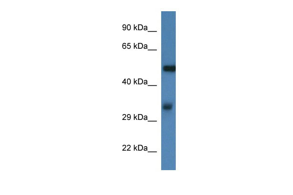
- 产品详情
- 实验流程
Application 
| WB |
|---|---|
| Primary Accession | Q61553 |
| Other Accession | NM_007984, NP_032010 |
| Reactivity | Human, Mouse, Rat, Zebrafish, Pig, Dog, Guinea Pig, Horse, Bovine |
| Predicted | Human, Mouse, Rat, Zebrafish, Pig, Chicken, Dog, Guinea Pig, Horse, Bovine |
| Host | Rabbit |
| Clonality | Polyclonal |
| Calculated MW | 54508 Da |
| Gene ID | 14086 |
|---|---|
| Alias Symbol | AI663989, Fan1, fascin-1 |
| Other Names | Fascin, Singed-like protein, Fscn1, Fan1, Snl |
| Format | Liquid. Purified antibody supplied in 1x PBS buffer with 0.09% (w/v) sodium azide and 2% sucrose. |
| Reconstitution & Storage | Add 50 ul of distilled water. Final anti-Fscn1 antibody concentration is 1 mg/ml in PBS buffer with 2% sucrose. For longer periods of storage, store at 20°C. Avoid repeat freeze-thaw cycles. |
| Precautions | Fscn1 antibody - N-terminal region is for research use only and not for use in diagnostic or therapeutic procedures. |
For Research Use Only. Not For Use In Diagnostic Procedures.
| Name | Fscn1 |
|---|---|
| Synonyms | Fan1, Snl |
| Function | Actin-binding protein that contains 2 major actin binding sites (By similarity). Organizes filamentous actin into parallel bundles (PubMed:7738015). Plays a role in the organization of actin filament bundles and the formation of microspikes, membrane ruffles, and stress fibers (By similarity). Important for the formation of a diverse set of cell protrusions, such as filopodia, and for cell motility and migration (PubMed:21685497). Mediates reorganization of the actin cytoskeleton and axon growth cone collapse in response to NGF (By similarity). |
| Cellular Location | Cytoplasm, cytosol {ECO:0000250|UniProtKB:Q16658}. Cytoplasm, cell cortex {ECO:0000250|UniProtKB:Q16658}. Cytoplasm, cytoskeleton {ECO:0000250|UniProtKB:Q16658}. Cytoplasm, cytoskeleton, stress fiber {ECO:0000250|UniProtKB:Q16658}. Cell projection, growth cone. Cell projection, filopodium. Cell projection, invadopodium. Cell projection, microvillus {ECO:0000250|UniProtKB:Q16658}. Cell junction {ECO:0000250|UniProtKB:Q16658}. Note=Colocalized with RUFY3 and F-actin at filipodia of the axonal growth cone (PubMed:24720729). Colocalized with DBN1 and F-actin at the transitional domain of the axonal growth cone (PubMed:24720729). {ECO:0000250|UniProtKB:Q16658, ECO:0000269|PubMed:24720729} |
| Tissue Location | Most abundant in brain. Detected at lower levels in lung, uterus, small intestine and spleen (at protein level) |
Research Areas
Application Protocols
Provided below are standard protocols that you may find useful for product applications.

终于等到您。ABCEPTA(百远生物)抗体产品。
点击下方“我要评价 ”按钮提交您的反馈信息,您的反馈和评价是我们最宝贵的财富之一,
我们将在1-3个工作日内处理您的反馈信息。
如有疑问,联系:0512-88856768 tech-china@abcepta.com.


