


 癌症的基本特征包括细胞增殖、血管生成、迁移、凋亡逃避机制和细胞永生等。找到癌症发生过程中这些通路的关键标记物和对应的抗体用于检测至关重要。
 为您推荐一个泛素化位点预测神器——泛素化分析工具,可以为您的蛋白的泛素化位点作出预测和评分。
 细胞自噬受体图形绘图工具为你的蛋白的细胞受体结合位点作出预测和评分,识别结合到自噬通路中的蛋白是非常重要的,便于让我们理解自噬在正常生理、病理过程中的作用,如发育、细胞分化、神经退化性疾病、压力条件下、感染和癌症。
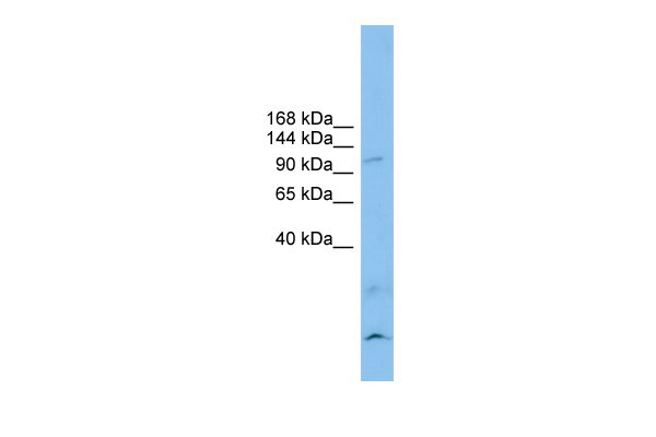
CLSTN3 antibody - N-terminal region
Rabbit Polyclonal Antibody
- 产品详情
- 实验流程
Application
| WB |
|---|---|
| Primary Accession | Q9BQT9 |
| Other Accession | NM_014718, NP_055533 |
| Reactivity | Human, Mouse, Rat, Rabbit, Pig, Dog, Guinea Pig, Horse, Bovine |
| Predicted | Human, Mouse, Rat, Chicken, Guinea Pig |
| Host | Rabbit |
| Clonality | Polyclonal |
| Calculated MW | 106098 Da |
| Gene ID | 9746 |
|---|---|
| Alias Symbol | CSTN3, KIAA0726, MGC131797, MGC138488, alcbeta, CDHR14 |
| Other Names | Calsyntenin-3, Alcadein-beta, Alc-beta, CLSTN3, CS3, KIAA0726 |
| Format | Liquid. Purified antibody supplied in 1x PBS buffer with 0.09% (w/v) sodium azide and 2% sucrose. |
| Reconstitution & Storage | Add 50 ul of distilled water. Final anti-CLSTN3 antibody concentration is 1 mg/ml in PBS buffer with 2% sucrose. For longer periods of storage, store at 20°C. Avoid repeat freeze-thaw cycles. |
| Precautions | CLSTN3 antibody - N-terminal region is for research use only and not for use in diagnostic or therapeutic procedures. |
| Name | CLSTN3 {ECO:0000303|PubMed:33101212, ECO:0000312|HGNC:HGNC:18371} |
|---|---|
| Function | Postsynaptic adhesion molecule that binds to presynaptic neurexins to mediate both excitatory and inhibitory synapse formation (PubMed:25352602). Promotes synapse development by acting as a cell adhesion molecule at the postsynaptic membrane, which associates with both neurexin-alpha and neurexin-beta proteins at the presynaptic membrane (PubMed:25352602). Regulates the balance between excitatory and inhibitory synapses by inhibiting formation of excitatory parallel- fiber synapses and promoting formation of inhibitory synapses in the same neuron (By similarity). May also be involved in ascorbate (vitamin C) uptake via its interaction with SLC23A2/SVCT2 (PubMed:34673103). Complex formation with APBA2 and APP, stabilizes APP metabolism and enhances APBA2-mediated suppression of beta-APP40 secretion, due to the retardation of intracellular APP maturation (Probable) (PubMed:12972431). |
| Cellular Location | Postsynaptic cell membrane {ECO:0000250|UniProtKB:Q99JH7}; Single-pass type I membrane protein. Endoplasmic reticulum membrane {ECO:0000250|UniProtKB:Q99JH7}; Single-pass type I membrane protein. Golgi apparatus membrane {ECO:0000250|UniProtKB:Q99JH7}; Single-pass type I membrane protein. Cell projection, dendrite {ECO:0000250|UniProtKB:Q99JH7}. Note=Most prominent in the postsynaptic specializations of asymmetric (type I) synapses with both axodendritic and axospinous localization {ECO:0000250|UniProtKB:Q99JH7} |
| Tissue Location | According to PubMed:12498782, expressed predominantly in the brain and in kidney (PubMed:12498782). Low levels in heart, skeletal muscle, liver, placenta, pancreas and lung (PubMed:12498782). According to PubMed:12972431, predominant expression in brain, and only marginal in kidney (PubMed:12972431). In brain, present throughout all cortical layers, highest levels in GABAergic neurons (based on morphology and distribution pattern) (PubMed:12972431). |
Research Areas
For Research Use Only. Not For Use In Diagnostic Procedures.
Application Protocols
Provided below are standard protocols that you may find useful for product applications.
终于等到您。ABCEPTA(百远生物)抗体产品。
点击下方“我要评价 ”按钮提交您的反馈信息,您的反馈和评价是我们最宝贵的财富之一,
我们将在1-3个工作日内处理您的反馈信息。
如有疑问,联系:0512-88856768 tech-china@abcepta.com.


