


 癌症的基本特征包括细胞增殖、血管生成、迁移、凋亡逃避机制和细胞永生等。找到癌症发生过程中这些通路的关键标记物和对应的抗体用于检测至关重要。
 为您推荐一个泛素化位点预测神器——泛素化分析工具,可以为您的蛋白的泛素化位点作出预测和评分。
 细胞自噬受体图形绘图工具为你的蛋白的细胞受体结合位点作出预测和评分,识别结合到自噬通路中的蛋白是非常重要的,便于让我们理解自噬在正常生理、病理过程中的作用,如发育、细胞分化、神经退化性疾病、压力条件下、感染和癌症。
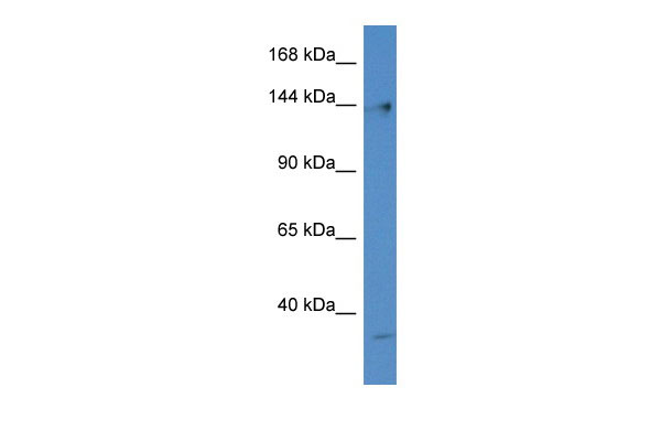
Polg antibody - N-terminal region
Rabbit Polyclonal Antibody
- 产品详情
- 实验流程
Application
| WB |
|---|---|
| Primary Accession | Q9QYV8 |
| Other Accession | NM_053528, NP_445980 |
| Reactivity | Human, Mouse, Rat, Zebrafish, Dog, Guinea Pig, Horse, Bovine, Yeast |
| Predicted | Human, Mouse, Rat, Zebrafish, Pig, Dog, Guinea Pig, Horse, Bovine |
| Host | Rabbit |
| Clonality | Polyclonal |
| Calculated MW | 136856 Da |
| Gene ID | 85472 |
|---|---|
| Other Names | DNA polymerase subunit gamma-1, 2.7.7.7, Mitochondrial DNA polymerase catalytic subunit, PolG-alpha, Polg, Mip1, Polg1 |
| Format | Liquid. Purified antibody supplied in 1x PBS buffer with 0.09% (w/v) sodium azide and 2% sucrose. |
| Reconstitution & Storage | Add 50 ul of distilled water. Final anti-Polg antibody concentration is 1 mg/ml in PBS buffer with 2% sucrose. For longer periods of storage, store at 20°C. Avoid repeat freeze-thaw cycles. |
| Precautions | Polg antibody - N-terminal region is for research use only and not for use in diagnostic or therapeutic procedures. |
| Name | Polg {ECO:0000312|RGD:620057} |
|---|---|
| Synonyms | Mip1, Polg1 |
| Function | Catalytic subunit of DNA polymerase gamma solely responsible for replication of mitochondrial DNA (mtDNA). Replicates both heavy and light strands of the circular mtDNA genome using a single-stranded DNA template, RNA primers and the four deoxyribonucleoside triphosphates as substrates (By similarity). Has 5' -> 3' polymerase activity. Functionally interacts with TWNK and SSBP1 at the replication fork to form a highly processive replisome, where TWNK unwinds the double- stranded DNA template prior to replication and SSBP1 covers the parental heavy strand to enable continuous replication of the entire mitochondrial genome. A single nucleotide incorporation cycle includes binding of the incoming nucleotide at the insertion site, a phosphodiester bond formation reaction that extends the 3'-end of the primer DNA, and translocation of the primer terminus to the post- insertion site. After completing replication of a mtDNA strand, mediates 3' -> 5' exonucleolytic degradation at the nick to enable proper ligation (By similarity). Highly accurate due to high nucleotide selectivity and 3' -> 5' exonucleolytic proofreading. Proficiently corrects base substitutions, single-base additions and deletions in non-repetitive sequences and short repeats, but displays lower proofreading activity when replicating longer homopolymeric stretches. Exerts exonuclease activity toward single-stranded DNA and double- stranded DNA containing 3'-terminal mispairs. When a misincorporation occurs, transitions from replication to a pro-nucleolytic editing mode and removes the missincorporated nucleoside in the exonuclease active site. Proceeds via an SN2 nucleolytic mechanism in which Asp-198 catalyzes phosphodiester bond hydrolysis and Glu-200 stabilizes the leaving group. As a result the primer strand becomes one nucleotide shorter and is positioned in the post-insertion site, ready to resume DNA synthesis (By similarity). Exerts 5'-deoxyribose phosphate (dRP) lyase activity and mediates repair-associated mtDNA synthesis (gap filling) in base-excision repair pathway. Catalyzes the release of the 5'-terminal 2-deoxyribose-5-phosphate sugar moiety from incised apurinic/apyrimidinic (AP) sites to produce a substrate for DNA ligase. The dRP lyase reaction does not require divalent metal ions and likely proceeds via a Schiff base intermediate in a beta-elimination reaction mechanism (By similarity). |
| Cellular Location | Mitochondrion {ECO:0000250|UniProtKB:P54098}. Mitochondrion matrix, mitochondrion nucleoid {ECO:0000250|UniProtKB:P54098} |
Research Areas
For Research Use Only. Not For Use In Diagnostic Procedures.
Application Protocols
Provided below are standard protocols that you may find useful for product applications.
终于等到您。ABCEPTA(百远生物)抗体产品。
点击下方“我要评价 ”按钮提交您的反馈信息,您的反馈和评价是我们最宝贵的财富之一,
我们将在1-3个工作日内处理您的反馈信息。
如有疑问,联系:0512-88856768 tech-china@abcepta.com.


