


 癌症的基本特征包括细胞增殖、血管生成、迁移、凋亡逃避机制和细胞永生等。找到癌症发生过程中这些通路的关键标记物和对应的抗体用于检测至关重要。
 为您推荐一个泛素化位点预测神器——泛素化分析工具,可以为您的蛋白的泛素化位点作出预测和评分。
 细胞自噬受体图形绘图工具为你的蛋白的细胞受体结合位点作出预测和评分,识别结合到自噬通路中的蛋白是非常重要的,便于让我们理解自噬在正常生理、病理过程中的作用,如发育、细胞分化、神经退化性疾病、压力条件下、感染和癌症。
FLII antibody - middle region
Rabbit Polyclonal Antibody
- 产品详情
- 实验流程
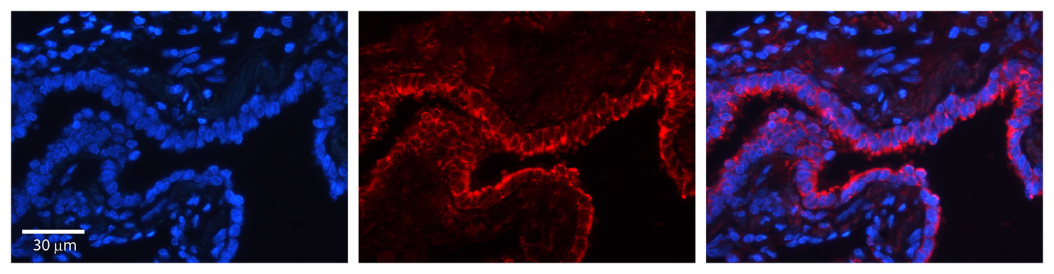
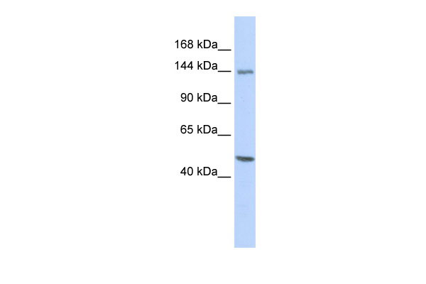
Application
| WB, IHC |
|---|---|
| Primary Accession | Q13045 |
| Other Accession | NM_002018, NP_002009 |
| Reactivity | Human, Mouse, Rat, Zebrafish, Dog, Guinea Pig, Horse, Bovine |
| Predicted | Human, Mouse, Rat, Zebrafish, Chicken, Dog, Guinea Pig, Horse, Bovine |
| Host | Rabbit |
| Clonality | Polyclonal |
| Calculated MW | 144751 Da |
| Gene ID | 2314 |
|---|---|
| Alias Symbol | FLI, FLIL, Fli1, MGC39265 |
| Other Names | Protein flightless-1 homolog, FLII, FLIL |
| Format | Liquid. Purified antibody supplied in 1x PBS buffer with 0.09% (w/v) sodium azide and 2% sucrose. |
| Reconstitution & Storage | Add 50 ul of distilled water. Final anti-FLII antibody concentration is 1 mg/ml in PBS buffer with 2% sucrose. For longer periods of storage, store at 20°C. Avoid repeat freeze-thaw cycles. |
| Precautions | FLII antibody - middle region is for research use only and not for use in diagnostic or therapeutic procedures. |
| Name | FLII |
|---|---|
| Synonyms | FLIL |
| Function | Is a regulator of actin polymerization, required for proper myofibril organization and regulation of the length of sarcomeric thin filaments (By similarity). It also plays a role in the assembly of cardiomyocyte cell adhesion complexes (By similarity). Regulates cytoskeletal rearrangements involved in cytokinesis and cell migration, by inhibiting Rac1-dependent paxillin phosphorylation (By similarity). May play a role as coactivator in transcriptional activation by hormone-activated nuclear receptors (NR) and acts in cooperation with NCOA2 and CARM1 (PubMed:14966289). Involved in estrogen hormone signaling. |
| Cellular Location | Nucleus {ECO:0000250|UniProtKB:Q9JJ28}. Cytoplasm, cytoskeleton {ECO:0000250|UniProtKB:Q9JJ28}. Cytoplasm, cytoskeleton, microtubule organizing center, centrosome {ECO:0000250|UniProtKB:Q9JJ28}. Cell projection, podosome {ECO:0000250|UniProtKB:Q9JJ28}. Cell junction, focal adhesion {ECO:0000250|UniProtKB:Q9JJ28}. Note=Colocalizes to actin-rich structures in blastocysts and, together with HRAS, RHOA and CDC42, in migrating fibroblasts. Localizes to centrosomes (By similarity) Localized to the core of macrophage podosomes (By similarity) {ECO:0000250|UniProtKB:Q9JJ28} |
| Tissue Location | Strongest expression in skeletal muscle with high expression also in the heart and lung. |
Research Areas
For Research Use Only. Not For Use In Diagnostic Procedures.
Application Protocols
Provided below are standard protocols that you may find useful for product applications.
REFERENCES
Campbell H.D.,et al.Genomics 42:46-54(1997).
Ota T.,et al.Nat. Genet. 36:40-45(2004).
Zody M.C.,et al.Nature 440:1045-1049(2006).
Gevaert K.,et al.Nat. Biotechnol. 21:566-569(2003).
Campbell H.D.,et al.Proc. Natl. Acad. Sci. U.S.A. 90:11386-11390(1993).
终于等到您。ABCEPTA(百远生物)抗体产品。
点击下方“我要评价 ”按钮提交您的反馈信息,您的反馈和评价是我们最宝贵的财富之一,
我们将在1-3个工作日内处理您的反馈信息。
如有疑问,联系:0512-88856768 tech-china@abcepta.com.


