


 癌症的基本特征包括细胞增殖、血管生成、迁移、凋亡逃避机制和细胞永生等。找到癌症发生过程中这些通路的关键标记物和对应的抗体用于检测至关重要。
 为您推荐一个泛素化位点预测神器——泛素化分析工具,可以为您的蛋白的泛素化位点作出预测和评分。
 细胞自噬受体图形绘图工具为你的蛋白的细胞受体结合位点作出预测和评分,识别结合到自噬通路中的蛋白是非常重要的,便于让我们理解自噬在正常生理、病理过程中的作用,如发育、细胞分化、神经退化性疾病、压力条件下、感染和癌症。
Opn3 antibody - C-terminal region
Rabbit Polyclonal Antibody
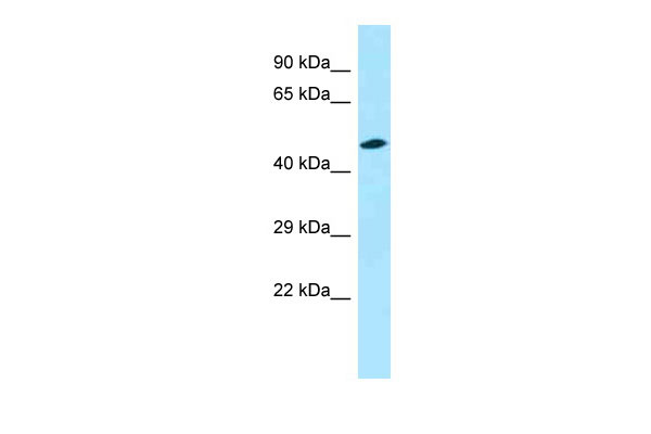
- 产品详情
- 实验流程
Application 
| WB |
|---|---|
| Primary Accession | Q9WUK7 |
| Other Accession | NM_010098, NP_034228 |
| Reactivity | Human, Mouse, Rat, Rabbit, Dog, Guinea Pig, Horse |
| Predicted | Human, Mouse, Rat, Rabbit, Chicken, Dog |
| Host | Rabbit |
| Clonality | Polyclonal |
| Calculated MW | 44947 Da |
| Gene ID | 13603 |
|---|---|
| Alias Symbol | ERO, Ecpn, MGC124138, panopsin |
| Other Names | Opsin-3, Encephalopsin, Panopsin, Opn3, Ecpn |
| Format | Liquid. Purified antibody supplied in 1x PBS buffer with 0.09% (w/v) sodium azide and 2% sucrose. |
| Reconstitution & Storage | Add 50 ul of distilled water. Final anti-Opn3 antibody concentration is 1 mg/ml in PBS buffer with 2% sucrose. For longer periods of storage, store at 20°C. Avoid repeat freeze-thaw cycles. |
| Precautions | Opn3 antibody - C-terminal region is for research use only and not for use in diagnostic or therapeutic procedures. |
For Research Use Only. Not For Use In Diagnostic Procedures.
| Name | Opn3 {ECO:0000312|MGI:MGI:1338022} |
|---|---|
| Synonyms | Ecpn |
| Function | G protein-coupled photoreceptor that selectively activates G(i/o) proteins in response to light, thereby decreasing intracellular cAMP levels (PubMed:30284927). Activation occurs when the opsin-bound cis-retinal chromophore absorbs a photon and isomerizes to all-trans- retinal, inducing a conformational change in the opsin that triggers a G protein-mediated phototransduction cascade. Mediates blue light- induced pigmentation in melanocytes by activating calcium/CAMK2 signaling, followed by phosphorylation of CREB, p38, ERK and MITF (By similarity). Required for TYR and DCT blue light-induced complex formation in melanocytes. Involved in keratinocyte differentiation in response to blue-light. Plays a role in melanocyte survival through regulation of intracellular calcium levels and subsequent BCL2/RAF1 signaling. Additionally, regulates apoptosis via cytochrome c release and subsequent activation of the caspase cascade. Responsible for ultraviolet A (UVA)-induced keratinocytes supranuclear melanin cap formation via activating calcium/CAMK2, CREB, and Akt signal transduction, and ultimately increasing DYNC1I1 and DCTN1 expression levels, thereby playing a role in the skin response to UV radiation. Required for the UVA-mediated induction of calcium and MAPK signaling resulting in the expression of MMP1, MMP2, MMP3, MMP9 and TIMP1 in dermal fibroblasts (By similarity). Also plays a role in light-mediated glucose uptake, mitochondrial respiration and fatty acid metabolism in brown adipocyte tissues (PubMed:32040503). May be involved in photorelaxation of airway smooth muscle cells, via blue light-dependent GPCR signaling pathways (PubMed:30284927). Also inhibits melanogenesis in melanocytes by specifically inhibiting MC1R-evoked cAMP signaling in a constitutive, light-independent manner (By similarity). In the paraventricular nucleus of the hypothalamus, inhibits MC4R-mediated cAMP signaling and activates KCNJ13 channel opening in a constitutive manner, thereby promoting food intake and modulating spontaneous firing of neurons (PubMed:39951488). |
| Cellular Location | Cell membrane {ECO:0000250|UniProtKB:Q9H1Y3}; Multi-pass membrane protein. Cytoplasm {ECO:0000250|UniProtKB:Q9H1Y3} |
| Tissue Location | Expressed in the eye (at protein level) (PubMed:30284927). Expressed in tracheal airway smooth muscle (PubMed:30284927). Expressed in brown adipocyte tissue; expression becomes more abundant during differentiation (PubMed:32040503) Strongly expressed in brain (PubMed:10234000). Highly expressed in the preoptic area and paraventricular nucleus of the hypothalamus (PubMed:10234000, PubMed:39951488). Shows highly patterned expression in other regions of the brain, being enriched in selected regions of the cerebral cortex, cerebellar Purkinje cells, a subset of striatal neurons, selected thalamic nuclei, and a subset of interneurons in the ventral horn of the spinal cord (PubMed:10234000) |
Research Areas
Application Protocols
Provided below are standard protocols that you may find useful for product applications.
REFERENCES
Blackshaw S.,et al.J. Neurosci. 19:3681-3690(1999).
Kasper G.,et al.Gene 295:27-32(2002).
Carninci P.,et al.Science 309:1559-1563(2005).

终于等到您。ABCEPTA(百远生物)抗体产品。
点击下方“我要评价 ”按钮提交您的反馈信息,您的反馈和评价是我们最宝贵的财富之一,
我们将在1-3个工作日内处理您的反馈信息。
如有疑问,联系:0512-88856768 tech-china@abcepta.com.


