


 癌症的基本特征包括细胞增殖、血管生成、迁移、凋亡逃避机制和细胞永生等。找到癌症发生过程中这些通路的关键标记物和对应的抗体用于检测至关重要。
 为您推荐一个泛素化位点预测神器——泛素化分析工具,可以为您的蛋白的泛素化位点作出预测和评分。
 细胞自噬受体图形绘图工具为你的蛋白的细胞受体结合位点作出预测和评分,识别结合到自噬通路中的蛋白是非常重要的,便于让我们理解自噬在正常生理、病理过程中的作用,如发育、细胞分化、神经退化性疾病、压力条件下、感染和癌症。
XPA antibody - C-terminal region
Rabbit Polyclonal Antibody
- 产品详情
- 实验流程
Application
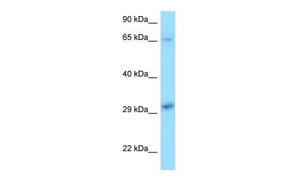
| WB |
|---|---|
| Primary Accession | P23025 |
| Other Accession | NM_000380, NP_000371 |
| Reactivity | Human, Mouse, Rat, Pig, Dog, Guinea Pig, Horse, Bovine |
| Predicted | Human, Mouse, Rat, Dog, Horse, Bovine |
| Host | Rabbit |
| Clonality | Polyclonal |
| Calculated MW | 31368 Da |
| Gene ID | 7507 |
|---|---|
| Alias Symbol | XP1, XPAC |
| Other Names | DNA repair protein complementing XP-A cells, Xeroderma pigmentosum group A-complementing protein, XPA, XPAC |
| Format | Liquid. Purified antibody supplied in 1x PBS buffer with 0.09% (w/v) sodium azide and 2% sucrose. |
| Reconstitution & Storage | Add 50 ul of distilled water. Final anti-XPA antibody concentration is 1 mg/ml in PBS buffer with 2% sucrose. For longer periods of storage, store at 20°C. Avoid repeat freeze-thaw cycles. |
| Precautions | XPA antibody - C-terminal region is for research use only and not for use in diagnostic or therapeutic procedures. |
| Name | XPA |
|---|---|
| Synonyms | XPAC |
| Function | Involved in DNA nucleotide excision repair (NER). Initiates repair by binding to damaged sites with various affinities, depending on the photoproduct and the transcriptional state of the region. Required for UV-induced CHEK1 phosphorylation and the recruitment of CEP164 to cyclobutane pyrimidine dimmers (CPD), sites of DNA damage after UV irradiation (PubMed:19197159). During NER stimulates the 5'-3' helicase activity of XPD/ERCC2 and the DNA translocase activity of XPB/ERCC3 (PubMed:31253769). Connects XPD/ERCC2 and XPB/ERCC3 during NER, retaining DNA near the XPB/ERCC3 active site, and stabilizing the complex in a different conformation than in transcribing TFIIH (PubMed:31253769). |
| Cellular Location | Nucleus |
| Tissue Location | Expressed in various cell lines and in skin fibroblasts. |
Research Areas
For Research Use Only. Not For Use In Diagnostic Procedures.
Application Protocols
Provided below are standard protocols that you may find useful for product applications.
REFERENCES
Tanaka K.,et al.Nature 348:73-76(1990).
Kalnine N.,et al.Submitted (MAY-2003) to the EMBL/GenBank/DDBJ databases.
Humphray S.J.,et al.Nature 429:369-374(2004).
Mural R.J.,et al.Submitted (JUL-2005) to the EMBL/GenBank/DDBJ databases.
Satokata I.,et al.Gene 136:345-348(1993).
终于等到您。ABCEPTA(百远生物)抗体产品。
点击下方“我要评价 ”按钮提交您的反馈信息,您的反馈和评价是我们最宝贵的财富之一,
我们将在1-3个工作日内处理您的反馈信息。
如有疑问,联系:0512-88856768 tech-china@abcepta.com.


