


 癌症的基本特征包括细胞增殖、血管生成、迁移、凋亡逃避机制和细胞永生等。找到癌症发生过程中这些通路的关键标记物和对应的抗体用于检测至关重要。
 为您推荐一个泛素化位点预测神器——泛素化分析工具,可以为您的蛋白的泛素化位点作出预测和评分。
 细胞自噬受体图形绘图工具为你的蛋白的细胞受体结合位点作出预测和评分,识别结合到自噬通路中的蛋白是非常重要的,便于让我们理解自噬在正常生理、病理过程中的作用,如发育、细胞分化、神经退化性疾病、压力条件下、感染和癌症。
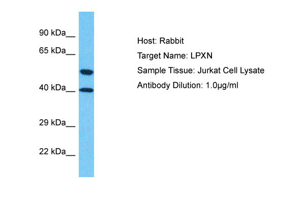
LPXN Antibody - C-terminal region
Rabbit Polyclonal Antibody
- 产品详情
- 实验流程
Application
| WB |
|---|---|
| Primary Accession | O60711 |
| Other Accession | NP_004802 |
| Reactivity | Human |
| Host | Rabbit |
| Clonality | Polyclonal |
| Calculated MW | 43332 Da |
| Gene ID | 9404 |
|---|---|
| Alias Symbol | LPXN, LDLP, |
| Other Names | Leupaxin, LPXN, LDLP |
| Format | Liquid. Purified antibody supplied in 1x PBS buffer with 0.09% (w/v) sodium azide and 2% sucrose. |
| Reconstitution & Storage | Add 50 &mu, l of distilled water. Final Anti-LPXN antibody concentration is 1 mg/ml in PBS buffer with 2% sucrose. For longer periods of storage, store at -20°C. Avoid repeat freeze-thaw cycles. |
| Precautions | LPXN Antibody - C-terminal region is for research use only and not for use in diagnostic or therapeutic procedures. |
| Name | LPXN |
|---|---|
| Synonyms | LDLP |
| Function | Transcriptional coactivator for androgen receptor (AR) and serum response factor (SRF). Contributes to the regulation of cell adhesion, spreading and cell migration and acts as a negative regulator in integrin-mediated cell adhesion events. Suppresses the integrin- induced tyrosine phosphorylation of paxillin (PXN). May play a critical role as an adapter protein in the formation of the adhesion zone in osteoclasts. Negatively regulates B-cell antigen receptor (BCR) signaling. |
| Cellular Location | Cytoplasm. Cell junction, focal adhesion. Nucleus. Cytoplasm, perinuclear region. Cell projection, podosome. Cell membrane. Note=Shuttles between the cytoplasm and nucleus. Recruited to the cell membrane following B-cell antigen receptor (BCR) cross-linking in B-cells. Enhanced focal adhesion kinase activity (PTK2/FAK) attenuates its nuclear accumulation and limits its ability to enhance serum response factor (SRF)-dependent gene transcription. Targeting to focal adhesions is essential for its tyrosine phosphorylation in response to bombesin |
| Tissue Location | Macrophages, monocytes and osteoclasts (at protein level). Strongly expressed in cells and tissues of hematopoietic origin. Highest expression in lymphoid tissues such as spleen, lymph node, thymus and appendix and in the vascular smooth muscle. Lower levels in bone marrow and fetal liver. Also expressed in peripheral blood lymphocytes and a number of hematopoietic cell lines. Very low levels found in epithelial cell lines. Expressed in prostate cancer (PCa) cells and its expression intensity is directly linked to PCa progression. |
Research Areas
For Research Use Only. Not For Use In Diagnostic Procedures.
Application Protocols
Provided below are standard protocols that you may find useful for product applications.
REFERENCES
Lipsky B.P.,et al.J. Biol. Chem. 273:11709-11713(1998).
Ota T.,et al.Nat. Genet. 36:40-45(2004).
Ebert L.,et al.Submitted (JUN-2004) to the EMBL/GenBank/DDBJ databases.
Suzuki Y.,et al.Submitted (APR-2005) to the EMBL/GenBank/DDBJ databases.
Taylor T.D.,et al.Nature 440:497-500(2006).
终于等到您。ABCEPTA(百远生物)抗体产品。
点击下方“我要评价 ”按钮提交您的反馈信息,您的反馈和评价是我们最宝贵的财富之一,
我们将在1-3个工作日内处理您的反馈信息。
如有疑问,联系:0512-88856768 tech-china@abcepta.com.


