


 癌症的基本特征包括细胞增殖、血管生成、迁移、凋亡逃避机制和细胞永生等。找到癌症发生过程中这些通路的关键标记物和对应的抗体用于检测至关重要。
 为您推荐一个泛素化位点预测神器——泛素化分析工具,可以为您的蛋白的泛素化位点作出预测和评分。
 细胞自噬受体图形绘图工具为你的蛋白的细胞受体结合位点作出预测和评分,识别结合到自噬通路中的蛋白是非常重要的,便于让我们理解自噬在正常生理、病理过程中的作用,如发育、细胞分化、神经退化性疾病、压力条件下、感染和癌症。
HOOK1 antibody - C-terminal region
Rabbit Polyclonal Antibody
- 产品详情
- 实验流程
Application
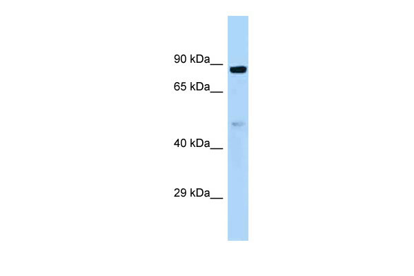
| WB |
|---|---|
| Primary Accession | Q9UJC3 |
| Other Accession | NM_015888, NP_056972 |
| Reactivity | Human, Mouse, Rat, Rabbit, Pig, Dog, Guinea Pig, Horse, Bovine |
| Predicted | Human, Mouse, Rat, Rabbit, Pig, Chicken, Dog, Guinea Pig, Horse, Bovine |
| Host | Rabbit |
| Clonality | Polyclonal |
| Calculated MW | 84648 Da |
| Gene ID | 51361 |
|---|---|
| Alias Symbol | HK1, MGC10642 |
| Other Names | Protein Hook homolog 1, h-hook1, hHK1, HOOK1 |
| Format | Liquid. Purified antibody supplied in 1x PBS buffer with 0.09% (w/v) sodium azide and 2% sucrose. |
| Reconstitution & Storage | Add 50 ul of distilled water. Final anti-HOOK1 antibody concentration is 1 mg/ml in PBS buffer with 2% sucrose. For longer periods of storage, store at 20°C. Avoid repeat freeze-thaw cycles. |
| Precautions | HOOK1 antibody - C-terminal region is for research use only and not for use in diagnostic or therapeutic procedures. |
| Name | HOOK1 (HGNC:19884) |
|---|---|
| Function | Component of the FTS/Hook/FHIP complex (FHF complex) (PubMed:18799622, PubMed:32073997). The FHF complex may function to promote vesicle trafficking and/or fusion via the homotypic vesicular protein sorting complex (the HOPS complex) (PubMed:18799622). FHF complex promotes the distribution of AP-4 complex to the perinuclear area of the cell (PubMed:32073997). Required for spermatid differentiation. Probably involved in the positioning of the microtubules of the manchette and the flagellum in relation to the membrane skeleton (By similarity). |
| Cellular Location | Cytoplasm. Cytoplasm, cytoskeleton. Note=Localizes to punctate cytoplasmic foci which do not appear to overlap with early or late endosomes, the endoplasmic reticulum, multivesicular bodies (MVBs), lysosomes, or mitochondria (By similarity). Often found in close association with microtubules (By similarity). Does not associate with the Golgi complex. During spermiogenesis, it localizes to the manchette in spermatids from steps 8-10. It is also present between the microtubule manchette and the nucleus. During manchette elongation, it is preferentially localized to the nuclear ring of the manchette, whereas the strong localization to the manchette decreases. In more mature spermatids, while the manchette migrates posteriorly, it localizes to punctuates spots. At later stages of spermatid differentiation, the punctuate expression pattern is found at both the attachment site and the proximal end of the elongated manchette. In contrast, it is not present in mature spermatozoa (By similarity) {ECO:0000250|UniProtKB:Q8BIL5} |
Research Areas
For Research Use Only. Not For Use In Diagnostic Procedures.
Application Protocols
Provided below are standard protocols that you may find useful for product applications.
REFERENCES
Kraemer H.,et al.Genetics 151:675-684(1999).
Ota T.,et al.Nat. Genet. 36:40-45(2004).
Gregory S.G.,et al.Nature 441:315-321(2006).
Mural R.J.,et al.Submitted (SEP-2005) to the EMBL/GenBank/DDBJ databases.
Walenta J.H.,et al.J. Cell Biol. 152:923-934(2001).
终于等到您。ABCEPTA(百远生物)抗体产品。
点击下方“我要评价 ”按钮提交您的反馈信息,您的反馈和评价是我们最宝贵的财富之一,
我们将在1-3个工作日内处理您的反馈信息。
如有疑问,联系:0512-88856768 tech-china@abcepta.com.


