


 癌症的基本特征包括细胞增殖、血管生成、迁移、凋亡逃避机制和细胞永生等。找到癌症发生过程中这些通路的关键标记物和对应的抗体用于检测至关重要。
 为您推荐一个泛素化位点预测神器——泛素化分析工具,可以为您的蛋白的泛素化位点作出预测和评分。
 细胞自噬受体图形绘图工具为你的蛋白的细胞受体结合位点作出预测和评分,识别结合到自噬通路中的蛋白是非常重要的,便于让我们理解自噬在正常生理、病理过程中的作用,如发育、细胞分化、神经退化性疾病、压力条件下、感染和癌症。
GPR37 Antibody - C-terminal region
Rabbit Polyclonal Antibody
- 产品详情
- 实验流程
Application
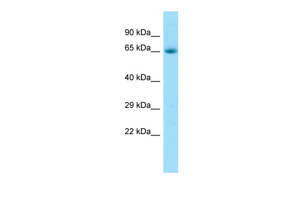
| WB |
|---|---|
| Primary Accession | O15354 |
| Other Accession | NM_005302, NP_005293 |
| Reactivity | Human, Mouse, Rat, Rabbit, Pig, Dog, Guinea Pig, Horse, Bovine |
| Predicted | Human, Mouse, Rat, Rabbit, Pig, Dog, Guinea Pig, Horse, Bovine |
| Host | Rabbit |
| Clonality | Polyclonal |
| Calculated MW | 67114 Da |
| Gene ID | 2861 |
|---|---|
| Alias Symbol | EDNRBL, PAELR, hET(B)R-LP |
| Other Names | Prosaposin receptor GPR37, Endothelin B receptor-like protein 1, ETBR-LP-1, G-protein coupled receptor 37, Parkin-associated endothelin receptor-like receptor, PAELR, GPR37 |
| Format | Liquid. Purified antibody supplied in 1x PBS buffer with 0.09% (w/v) sodium azide and 2% sucrose. |
| Reconstitution & Storage | Add 50 ul of distilled water. Final anti-GPR37 antibody concentration is 1 mg/ml in PBS buffer with 2% sucrose. For longer periods of storage, store at 20°C. Avoid repeat freeze-thaw cycles. |
| Precautions | GPR37 Antibody - C-terminal region is for research use only and not for use in diagnostic or therapeutic procedures. |
| Name | GPR37 |
|---|---|
| Function | G-protein-coupled receptor that plays a role in several physiological pathways such as resolution of inflammatory pain and oligodendrocyte differentiation (By similarity). Acts as a receptor for several ligands including prosaposin, osteocalcin or neuroprotectin D1. Ligand binding induces endocytosis, followed by an ERK phosphorylation cascade (PubMed:11439185, PubMed:23690594). Acts as a receptor for osteocalcin (OCN) to regulate oligodendrocyte differentiation and central nervous system myelination. Mechanistically, plays a negative role in oligodendrocyte differentiation and myelination during development via activation of the ERK1/2 signaling pathway. Therefore, regulates the stability of myelin or resistance of myelin itself to demyelination. Upon activation by neuroprotectin D1 (NPD1), promotes the activation of phagocytosis in macrophages as well as the shift in cytokine release toward an anti-inflammatory profile, and thus helps to reverse inflammatory pain. In addition, the increased macrophage phagocytosis mediates protection against sepsis upon pathogen infection. Additionally, extracellular vesicles derived from efferocyte express prosaposin, which binds to macrophage GPR37 to increase expression of the efferocytosis receptor TIM4 via an ERK-AP1-dependent signaling axis, leading to increased macrophage efferocytosis efficiency and accelerated resolution of inflammation (By similarity). May also act as a maturation factor of LRP6, protecting LRP6 from the endoplasmic reticulum (ER)-associated protein degradation (ERAD) and thereby promoting the Wnt/beta-catenin signaling pathway (PubMed:28341812). |
| Cellular Location | Cell projection, dendrite. Synapse Cell membrane; Multi-pass membrane protein. Endoplasmic reticulum membrane; Multi-pass membrane protein |
| Tissue Location | Expressed in brain and spinal cord, and at lower levels in testis, placenta and liver, but no detectable expression observed in any other tissue. When overexpressed in cells, tends to become insoluble and unfolded. Accumulation of the unfolded protein may lead to dopaminergic neuronal death in juvenile Parkinson disease (PDJ). |
Research Areas
For Research Use Only. Not For Use In Diagnostic Procedures.
Application Protocols
Provided below are standard protocols that you may find useful for product applications.
REFERENCES
Marazziti D.,et al.Genomics 45:68-77(1997).
Donohue P.J.,et al.Brain Res. Mol. Brain Res. 54:152-160(1998).
Zeng Z.,et al.Biochem. Biophys. Res. Commun. 233:559-567(1997).
Imai Y.,et al.Cell 105:891-902(2001).
Hillier L.W.,et al.Nature 424:157-164(2003).
终于等到您。ABCEPTA(百远生物)抗体产品。
点击下方“我要评价 ”按钮提交您的反馈信息,您的反馈和评价是我们最宝贵的财富之一,
我们将在1-3个工作日内处理您的反馈信息。
如有疑问,联系:0512-88856768 tech-china@abcepta.com.


