


 癌症的基本特征包括细胞增殖、血管生成、迁移、凋亡逃避机制和细胞永生等。找到癌症发生过程中这些通路的关键标记物和对应的抗体用于检测至关重要。
 为您推荐一个泛素化位点预测神器——泛素化分析工具,可以为您的蛋白的泛素化位点作出预测和评分。
 细胞自噬受体图形绘图工具为你的蛋白的细胞受体结合位点作出预测和评分,识别结合到自噬通路中的蛋白是非常重要的,便于让我们理解自噬在正常生理、病理过程中的作用,如发育、细胞分化、神经退化性疾病、压力条件下、感染和癌症。
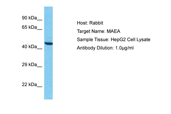
MAEA Antibody - N-terminal region
Rabbit Polyclonal Antibody
- 产品详情
- 实验流程
Application
| WB |
|---|---|
| Primary Accession | Q7L5Y9 |
| Reactivity | Human |
| Host | Rabbit |
| Clonality | Polyclonal |
| Calculated MW | 45287 Da |
| Gene ID | 10296 |
|---|---|
| Alias Symbol | MAEA, EMP, HLC10, PIG5, |
| Other Names | Macrophage erythroblast attacher, Cell proliferation-inducing gene 5 protein, Erythroblast macrophage protein, Human lung cancer oncogene 10 protein, HLC-10, MAEA, EMP |
| Format | Liquid. Purified antibody supplied in 1x PBS buffer with 0.09% (w/v) sodium azide and 2% sucrose. |
| Reconstitution & Storage | Add 50 &mu, l of distilled water. Final Anti-MAEA antibody concentration is 1 mg/ml in PBS buffer with 2% sucrose. For longer periods of storage, store at -20°C. Avoid repeat freeze-thaw cycles. |
| Precautions | MAEA Antibody - N-terminal region is for research use only and not for use in diagnostic or therapeutic procedures. |
| Name | MAEA |
|---|---|
| Function | Core component of the CTLH E3 ubiquitin-protein ligase complex that selectively accepts ubiquitin from UBE2H and mediates ubiquitination and subsequent proteasomal degradation of the transcription factor HBP1. MAEA and RMND5A are both required for catalytic activity of the CTLH E3 ubiquitin-protein ligase complex (PubMed:29911972). MAEA is required for normal cell proliferation (PubMed:29911972). The CTLH E3 ubiquitin-protein ligase complex is not required for the degradation of enzymes involved in gluconeogenesis, such as FBP1 (PubMed:29911972). Plays a role in erythroblast enucleation during erythrocyte maturation and in the development of mature macrophages (By similarity). Mediates the attachment of erythroid cell to mature macrophages; this MAEA-mediated contact inhibits erythroid cell apoptosis (PubMed:9763581). Participates in erythroblastic island formation, which is the functional unit of definitive erythropoiesis. Associates with F-actin to regulate actin distribution in erythroblasts and macrophages (By similarity). May contribute to nuclear architecture and cells division events (Probable). |
| Cellular Location | Cytoplasm {ECO:0000250|UniProtKB:Q4VC33}. Nucleus, nucleoplasm. Nucleus matrix. Cell membrane. Cytoplasm, cytoskeleton. Note=Detected in a nuclear, speckled- like pattern (PubMed:16510120). Localized with condensed chromatin at prophase; Detected in nuclear spindle poles at metaphase and in the contractile ring during telophase and cytokinesis (PubMed:16510120) Present in cytoplasm, nuclear matrix and at the cell surface in macrophages; predominantly nuclear in immature macrophages and predominantly detected at the cell surface in mature macrophages Colocalizes with F-actin in macrophages (By similarity) {ECO:0000250|UniProtKB:Q4VC33, ECO:0000269|PubMed:16510120} |
| Tissue Location | Detected at macrophage membranes (at protein level). Ubiquitous. |
Research Areas
For Research Use Only. Not For Use In Diagnostic Procedures.
Application Protocols
Provided below are standard protocols that you may find useful for product applications.
REFERENCES
Kim J.W.,et al.Submitted (FEB-2003) to the EMBL/GenBank/DDBJ databases.
Ota T.,et al.Nat. Genet. 36:40-45(2004).
Hanspal M.,et al.Blood 92:2940-2950(1998).
Kim J.W.,et al.Submitted (NOV-2002) to the EMBL/GenBank/DDBJ databases.
Kalnine N.,et al.Submitted (MAY-2003) to the EMBL/GenBank/DDBJ databases.
终于等到您。ABCEPTA(百远生物)抗体产品。
点击下方“我要评价 ”按钮提交您的反馈信息,您的反馈和评价是我们最宝贵的财富之一,
我们将在1-3个工作日内处理您的反馈信息。
如有疑问,联系:0512-88856768 tech-china@abcepta.com.


