


 癌症的基本特征包括细胞增殖、血管生成、迁移、凋亡逃避机制和细胞永生等。找到癌症发生过程中这些通路的关键标记物和对应的抗体用于检测至关重要。
 为您推荐一个泛素化位点预测神器——泛素化分析工具,可以为您的蛋白的泛素化位点作出预测和评分。
 细胞自噬受体图形绘图工具为你的蛋白的细胞受体结合位点作出预测和评分,识别结合到自噬通路中的蛋白是非常重要的,便于让我们理解自噬在正常生理、病理过程中的作用,如发育、细胞分化、神经退化性疾病、压力条件下、感染和癌症。
NLRX1 Antibody - C-terminal region
Rabbit Polyclonal Antibody
- 产品详情
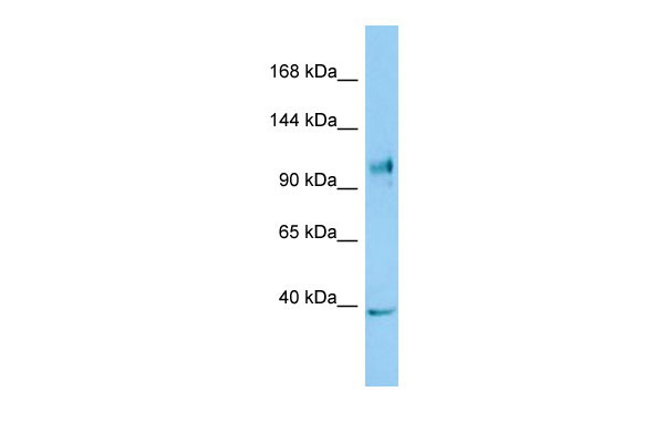
- 实验流程
Application
| WB |
|---|---|
| Primary Accession | Q86UT6 |
| Other Accession | NM_024618, NP_078894 |
| Reactivity | Human, Mouse, Rat, Rabbit, Pig, Dog, Guinea Pig, Horse, Bovine |
| Predicted | Human, Mouse, Rat, Rabbit, Pig, Dog, Guinea Pig, Horse, Bovine |
| Host | Rabbit |
| Clonality | Polyclonal |
| Calculated MW | 107616 Da |
| Gene ID | 79671 |
|---|---|
| Alias Symbol | CLR11.3, DLNB26, FLJ21478, MGC131937, MGC21025, NOD26, NOD5, NOD9 |
| Other Names | NLR family member X1, Caterpiller protein 11.3, CLR11.3, Nucleotide-binding oligomerization domain protein 26, Nucleotide-binding oligomerization domain protein 5, Nucleotide-binding oligomerization domain protein 9, NLRX1, NOD26, NOD5, NOD9 |
| Format | Liquid. Purified antibody supplied in 1x PBS buffer with 0.09% (w/v) sodium azide and 2% sucrose. |
| Reconstitution & Storage | Add 50 ul of distilled water. Final anti-NLRX1 antibody concentration is 1 mg/ml in PBS buffer with 2% sucrose. For longer periods of storage, store at 20°C. Avoid repeat freeze-thaw cycles. |
| Precautions | NLRX1 Antibody - C-terminal region is for research use only and not for use in diagnostic or therapeutic procedures. |
| Name | NLRX1 |
|---|---|
| Function | Participates in antiviral signaling. Acts as a negative regulator of MAVS-mediated antiviral responses, through the inhibition of the virus-induced RLH (RIG-like helicase)-MAVS interaction (PubMed:18200010). Instead, promotes autophagy by interacting with TUFM and subsequently recruiting the autophagy-related proteins ATG5 and ATG12 (PubMed:22749352). Also regulates MAVS-dependent NLRP3 inflammasome activation to attenuate apoptosis (PubMed:27393910). Has no inhibitory function on NF-kappa-B signaling pathway, but enhances NF-kappa-B and JUN N-terminal kinase dependent signaling through the production of reactive oxygen species (PubMed:18219313). Regulates viral mediated-inflammation and energy metabolism in a sex-dependent manner (By similarity). In females, prevents uncontrolled inflammation and energy metabolism and thus, may contribute to the sex differences observed in infectious and inflammatory diseases (By similarity). |
| Cellular Location | Mitochondrion outer membrane |
| Tissue Location | Ubiquitously expressed. Strongest expression in mammary gland, heart and muscle. Detected in HeLa, HEK293T, THP-1, HL- 60, Raji and Jurkat cell lines (at protein level) |
Research Areas
For Research Use Only. Not For Use In Diagnostic Procedures.
Application Protocols
Provided below are standard protocols that you may find useful for product applications.
REFERENCES
Inohara N.,et al.Nat. Rev. Immunol. 3:371-382(2003).
Kronos K.,et al.Submitted (FEB-2007) to the EMBL/GenBank/DDBJ databases.
Kubo T.,et al.Submitted (OCT-2002) to the EMBL/GenBank/DDBJ databases.
Ota T.,et al.Nat. Genet. 36:40-45(2004).
Inohara N.,et al.Annu. Rev. Biochem. 74:355-383(2005).
终于等到您。ABCEPTA(百远生物)抗体产品。
点击下方“我要评价 ”按钮提交您的反馈信息,您的反馈和评价是我们最宝贵的财富之一,
我们将在1-3个工作日内处理您的反馈信息。
如有疑问,联系:0512-88856768 tech-china@abcepta.com.


