


 癌症的基本特征包括细胞增殖、血管生成、迁移、凋亡逃避机制和细胞永生等。找到癌症发生过程中这些通路的关键标记物和对应的抗体用于检测至关重要。
 为您推荐一个泛素化位点预测神器——泛素化分析工具,可以为您的蛋白的泛素化位点作出预测和评分。
 细胞自噬受体图形绘图工具为你的蛋白的细胞受体结合位点作出预测和评分,识别结合到自噬通路中的蛋白是非常重要的,便于让我们理解自噬在正常生理、病理过程中的作用,如发育、细胞分化、神经退化性疾病、压力条件下、感染和癌症。
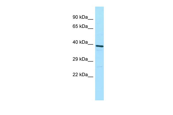
ATP6V0D1 Antibody - C-terminal region
Rabbit Polyclonal Antibody
- 产品详情
- 实验流程
Application
| WB |
|---|---|
| Primary Accession | P61421 |
| Other Accession | NM_004691, NP_004682 |
| Reactivity | Human, Mouse, Rat, Rabbit, Zebrafish, Pig, Dog, Guinea Pig, Horse, Bovine, Yeast |
| Predicted | Human, Mouse, Rat, Rabbit, Zebrafish, Pig, Dog, Guinea Pig, Horse, Bovine, Yeast |
| Host | Rabbit |
| Clonality | Polyclonal |
| Calculated MW | 40329 Da |
| Gene ID | 9114 |
|---|---|
| Alias Symbol | ATP6D, ATP6DV, FLJ43534, P39, VATX, VMA6, VPATPD |
| Other Names | V-type proton ATPase subunit d 1, V-ATPase subunit d 1, 32 kDa accessory protein, V-ATPase 40 kDa accessory protein, V-ATPase AC39 subunit, p39, Vacuolar proton pump subunit d 1, ATP6V0D1, ATP6D, VPATPD |
| Format | Liquid. Purified antibody supplied in 1x PBS buffer with 0.09% (w/v) sodium azide and 2% sucrose. |
| Reconstitution & Storage | Add 50 ul of distilled water. Final anti-ATP6V0D1 antibody concentration is 1 mg/ml in PBS buffer with 2% sucrose. For longer periods of storage, store at 20°C. Avoid repeat freeze-thaw cycles. |
| Precautions | ATP6V0D1 Antibody - C-terminal region is for research use only and not for use in diagnostic or therapeutic procedures. |
| Name | ATP6V0D1 (HGNC:13724) |
|---|---|
| Synonyms | ATP6D, VPATPD |
| Function | Subunit of the V0 complex of vacuolar(H+)-ATPase (V-ATPase), a multisubunit enzyme composed of a peripheral complex (V1) that hydrolyzes ATP and a membrane integral complex (V0) that translocates protons (PubMed:28296633, PubMed:30374053, PubMed:33065002). V-ATPase is responsible for acidifying and maintaining the pH of intracellular compartments and in some cell types, is targeted to the plasma membrane, where it is responsible for acidifying the extracellular environment (PubMed:30374053). May play a role in coupling of proton transport and ATP hydrolysis (By similarity). In aerobic conditions, involved in intracellular iron homeostasis, thus triggering the activity of Fe(2+) prolyl hydroxylase (PHD) enzymes, and leading to HIF1A hydroxylation and subsequent proteasomal degradation (PubMed:28296633). May play a role in cilium biogenesis through regulation of the transport and the localization of proteins to the cilium (By similarity). |
| Cellular Location | Membrane; Peripheral membrane protein; Cytoplasmic side. Lysosome membrane; Peripheral membrane protein. Cytoplasmic vesicle, clathrin-coated vesicle membrane {ECO:0000250|UniProtKB:P61420}; Peripheral membrane protein. Note=Localizes to centrosome and the base of the cilium {ECO:0000250|UniProtKB:Q6PGV1} |
| Tissue Location | Ubiquitous. |
Research Areas
For Research Use Only. Not For Use In Diagnostic Procedures.
Application Protocols
Provided below are standard protocols that you may find useful for product applications.
REFERENCES
van Hille B.,et al.Biochem. Biophys. Res. Commun. 197:15-21(1993).
Agarwal A.K.,et al.Biochem. Biophys. Res. Commun. 279:543-547(2000).
Bhat K.S.,et al.Submitted (NOV-1992) to the EMBL/GenBank/DDBJ databases.
Smith A.N.,et al.Gene 297:169-177(2002).
Burkard T.R.,et al.BMC Syst. Biol. 5:17-17(2011).
终于等到您。ABCEPTA(百远生物)抗体产品。
点击下方“我要评价 ”按钮提交您的反馈信息,您的反馈和评价是我们最宝贵的财富之一,
我们将在1-3个工作日内处理您的反馈信息。
如有疑问,联系:0512-88856768 tech-china@abcepta.com.


