


 癌症的基本特征包括细胞增殖、血管生成、迁移、凋亡逃避机制和细胞永生等。找到癌症发生过程中这些通路的关键标记物和对应的抗体用于检测至关重要。
 为您推荐一个泛素化位点预测神器——泛素化分析工具,可以为您的蛋白的泛素化位点作出预测和评分。
 细胞自噬受体图形绘图工具为你的蛋白的细胞受体结合位点作出预测和评分,识别结合到自噬通路中的蛋白是非常重要的,便于让我们理解自噬在正常生理、病理过程中的作用,如发育、细胞分化、神经退化性疾病、压力条件下、感染和癌症。
B4GALNT2 Antibody - N-terminal region
Rabbit Polyclonal Antibody
- 产品详情
- 实验流程
Application
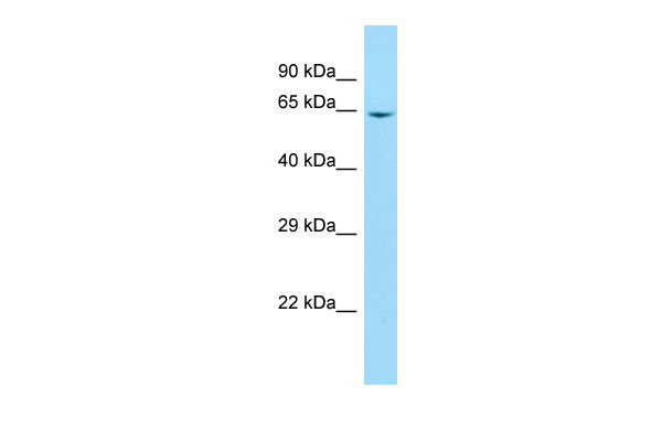
| WB |
|---|---|
| Primary Accession | Q8NHY0 |
| Other Accession | NM_153446, NP_703147 |
| Reactivity | Human |
| Predicted | Human |
| Host | Rabbit |
| Clonality | Polyclonal |
| Calculated MW | 63258 Da |
| Gene ID | 124872 |
|---|---|
| Alias Symbol | B4GALT, GALGT2, MGC142235, MGC142237 |
| Other Names | Beta-1, 4 N-acetylgalactosaminyltransferase 2, 2.4.1.-, Sd(a) beta-1, 4-GalNAc transferase, UDP-GalNAc:Neu5Aca2-3Galb-R b1, 4-N-acetylgalactosaminyltransferase, B4GALNT2, GALGT2 |
| Format | Liquid. Purified antibody supplied in 1x PBS buffer with 0.09% (w/v) sodium azide and 2% sucrose. |
| Reconstitution & Storage | Add 50 ul of distilled water. Final anti-B4GALNT2 antibody concentration is 1 mg/ml in PBS buffer with 2% sucrose. For longer periods of storage, store at 20°C. Avoid repeat freeze-thaw cycles. |
| Precautions | B4GALNT2 Antibody - N-terminal region is for research use only and not for use in diagnostic or therapeutic procedures. |
| Name | B4GALNT2 {ECO:0000303|PubMed:30067891, ECO:0000312|HGNC:HGNC:24136} |
|---|---|
| Function | Beta-1,4 N-acetylgalactosaminyltransferase involved in the biosynthesis of Sd(a) histo-blood group antigen. Catalyzes the transfer of N-acetylgalactosamine (GalNAc) group in a beta-1,4-linkage from UDP- GalNAc to the galactose residue of NeuAcalpha2->3Gal-R to form Sd(a) glycan epitope GalNAcbeta1->4(NeuAcalpha2->3)Gal-R. The Sd(a) epitope is carried in O- and N-linked glycoproteins and glycolipids, including O-linked core 1 structures on GYPA/glycophorin, SLC4A1 and SLC29A1 in erythrocytes, N-linked glycans attached to the Tamm-Horsfall glycoprotein UMOD/uromodulin in renal fluids, O-linked core 3 glycans on mucins in colon and neolactosides in gastric mucosa (PubMed:12678917, PubMed:14688233, PubMed:16024623, PubMed:35409292). Confers protection against influenza A virus strains that attach to NeuAcalpha2->3-carrying host receptors. Modifies N-glycan chains on host receptors and prevents virus entry into cells (PubMed:28813663). |
| Cellular Location | [Isoform 1]: Golgi apparatus, trans-Golgi network membrane; Single-pass type II membrane protein. Cytoplasmic vesicle membrane; Single-pass type II membrane protein. Note=Partially colocalizes with EEA1 and LAMP2 in early endosomes and lysosomes, respectively. |
| Tissue Location | Widely expressed. Highly expressed in colon and to a lesser extent in kidney, stomach, ileum and rectum |
Research Areas
For Research Use Only. Not For Use In Diagnostic Procedures.
Application Protocols
Provided below are standard protocols that you may find useful for product applications.
REFERENCES
Montiel M.D.,et al.Biochem. J. 373:369-379(2003).
Lo Presti L.,et al.J. Biochem. 134:675-682(2003).
Ota T.,et al.Nat. Genet. 36:40-45(2004).
Zody M.C.,et al.Nature 440:1045-1049(2006).
Mural R.J.,et al.Submitted (SEP-2005) to the EMBL/GenBank/DDBJ databases.
终于等到您。ABCEPTA(百远生物)抗体产品。
点击下方“我要评价 ”按钮提交您的反馈信息,您的反馈和评价是我们最宝贵的财富之一,
我们将在1-3个工作日内处理您的反馈信息。
如有疑问,联系:0512-88856768 tech-china@abcepta.com.


