


 癌症的基本特征包括细胞增殖、血管生成、迁移、凋亡逃避机制和细胞永生等。找到癌症发生过程中这些通路的关键标记物和对应的抗体用于检测至关重要。
 为您推荐一个泛素化位点预测神器——泛素化分析工具,可以为您的蛋白的泛素化位点作出预测和评分。
 细胞自噬受体图形绘图工具为你的蛋白的细胞受体结合位点作出预测和评分,识别结合到自噬通路中的蛋白是非常重要的,便于让我们理解自噬在正常生理、病理过程中的作用,如发育、细胞分化、神经退化性疾病、压力条件下、感染和癌症。
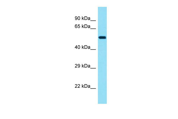
KRT78 Antibody - middle region
Rabbit Polyclonal Antibody
- 产品详情
- 实验流程
Application
| WB |
|---|---|
| Primary Accession | Q8N1N4 |
| Other Accession | NM_173352, NP_775487 |
| Reactivity | Human, Mouse, Rat, Rabbit, Pig, Dog, Guinea Pig, Bovine |
| Predicted | Human, Mouse, Rat, Rabbit, Pig, Dog, Guinea Pig, Bovine |
| Host | Rabbit |
| Clonality | Polyclonal |
| Calculated MW | 56866 Da |
| Gene ID | 196374 |
|---|---|
| Alias Symbol | K5B, Kb40 |
| Other Names | Keratin, type II cytoskeletal 78, Cytokeratin-78, CK-78, Keratin-5b, Keratin-78, K78, Type-II keratin Kb40, KRT78, K5B, KB40 |
| Format | Liquid. Purified antibody supplied in 1x PBS buffer with 0.09% (w/v) sodium azide and 2% sucrose. |
| Reconstitution & Storage | Add 50 ul of distilled water. Final anti-KRT78 antibody concentration is 1 mg/ml in PBS buffer with 2% sucrose. For longer periods of storage, store at 20°C. Avoid repeat freeze-thaw cycles. |
| Precautions | KRT78 Antibody - middle region is for research use only and not for use in diagnostic or therapeutic procedures. |
| Name | KRT78 |
|---|---|
| Synonyms | K5B, KB40 |
| Tissue Location | In non-keratinising esophageal and vaginal epithelium, strongly expressed in the basal and parabasal/lower suprabasal cell layers with considerably decreased expression in the mid/upper suprabasal layers (at protein level) (PubMed:26340985). A similar gradient from basal to lower suprabasal layers is seen in the partially keratinised dorsal tongue epithelium, in the scalp and in the plantar epidermis (at protein level) (PubMed:26340985). Extension of expression into the suprabasal compartments is distinctly more pronounced in non-keratinising epithelia than in keratinising epithelia and epidermis (at protein level) (PubMed:26340985). In scalp sections, present in the interfollicular epidermis and infundibulum including the entire outer root sheath of the hair follicles and also in the sebocytes (at protein level) (PubMed:26340985). In sweat glands, expressed in peripheral and luminal cells of the lower duct and in peripheral cells of the middle/upper duct with no expression observed in luminal cells (at protein level) (PubMed:26340985). In embryos at the 14th week of pregnancy, detected in basal and parabasal layers but is absent from the uppermost epidermal layer (at protein level) (PubMed:26340985). Expressed in tongue epithelium (PubMed:15737194) |
Research Areas
For Research Use Only. Not For Use In Diagnostic Procedures.
Application Protocols
Provided below are standard protocols that you may find useful for product applications.
REFERENCES
Ota T.,et al.Nat. Genet. 36:40-45(2004).
Bechtel S.,et al.BMC Genomics 8:399-399(2007).
Scherer S.E.,et al.Nature 440:346-351(2006).
Mural R.J.,et al.Submitted (JUL-2005) to the EMBL/GenBank/DDBJ databases.
Hesse M.,et al.J. Cell Sci. 114:2569-2575(2001).
终于等到您。ABCEPTA(百远生物)抗体产品。
点击下方“我要评价 ”按钮提交您的反馈信息,您的反馈和评价是我们最宝贵的财富之一,
我们将在1-3个工作日内处理您的反馈信息。
如有疑问,联系:0512-88856768 tech-china@abcepta.com.


