


 癌症的基本特征包括细胞增殖、血管生成、迁移、凋亡逃避机制和细胞永生等。找到癌症发生过程中这些通路的关键标记物和对应的抗体用于检测至关重要。
 为您推荐一个泛素化位点预测神器——泛素化分析工具,可以为您的蛋白的泛素化位点作出预测和评分。
 细胞自噬受体图形绘图工具为你的蛋白的细胞受体结合位点作出预测和评分,识别结合到自噬通路中的蛋白是非常重要的,便于让我们理解自噬在正常生理、病理过程中的作用,如发育、细胞分化、神经退化性疾病、压力条件下、感染和癌症。
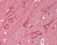
Anti-CLASP2 Antibody (N-Terminus, clone KT69)
Rat Anti Mouse Monoclonal Antibody
- 产品详情
- 实验流程
Application
| WB, IHC-P, IF |
|---|---|
| Primary Accession | O75122 |
| Predicted | Human, Mouse |
| Host | Rat |
| Clonality | Monoclonal |
| Isotype | IgG2b |
| Clone Names | KT69 |
| Calculated MW | 141064 Da |
| Concentration (mg/ml) | 1 mg/ml |
| Gene ID | 23122 |
|---|---|
| Alias Symbol | CLASP2 |
| Other Names | CLASP2, CLIP-associating protein 2, Protein Orbit homolog 2, HOrbit2, KIAA0627 |
| Target/Specificity | Mouse CLASP2 |
| Reconstitution & Storage | PBS, 0.1% sodium azide. Short term 4°C, long term aliquot and store at -20°C, avoid freeze thaw cycles. Store undiluted. |
| Precautions | Anti-CLASP2 Antibody (N-Terminus, clone KT69) is for research use only and not for use in diagnostic or therapeutic procedures. |
| Name | CLASP2 |
|---|---|
| Synonyms | KIAA0627 |
| Function | Microtubule plus-end tracking protein that promotes the stabilization of dynamic microtubules (PubMed:26003921). Involved in the nucleation of noncentrosomal microtubules originating from the trans-Golgi network (TGN). Required for the polarization of the cytoplasmic microtubule arrays in migrating cells towards the leading edge of the cell. May act at the cell cortex to enhance the frequency of rescue of depolymerizing microtubules by attaching their plus-ends to cortical platforms composed of ERC1 and PHLDB2 (PubMed:16824950). This cortical microtubule stabilizing activity is regulated at least in part by phosphatidylinositol 3-kinase signaling. Also performs a similar stabilizing function at the kinetochore which is essential for the bipolar alignment of chromosomes on the mitotic spindle (PubMed:16866869, PubMed:16914514). Acts as a mediator of ERBB2- dependent stabilization of microtubules at the cell cortex. |
| Cellular Location | Cytoplasm, cytoskeleton. Cytoplasm, cytoskeleton, microtubule organizing center, centrosome. Chromosome, centromere, kinetochore. Cytoplasm, cytoskeleton, spindle. Golgi apparatus {ECO:0000250|UniProtKB:Q8BRT1}. Golgi apparatus, trans-Golgi network. Cell membrane. Cell projection, ruffle membrane. Cytoplasm, cell cortex. Note=Localizes to microtubule plus ends (PubMed:15631994). Localizes to centrosomes, kinetochores and the mitotic spindle from prometaphase. Subsequently localizes to the spindle midzone from anaphase and to the midbody from telophase (PubMed:16866869, PubMed:16914514). In migrating cells localizes to the plus ends of microtubules within the cell body and to the entire microtubule lattice within the lamella. Localizes to the cell cortex and this requires ERC1 and PHLDB2 (PubMed:16824950). Colocalizes with KANK1 at the cell cortex, likely recruited in cortical microtubule stabilization complexes (CMSC) at focal adhesions rims (PubMed:27410476). The MEMO1-RHOA-DIAPH1 signaling pathway controls localization of the phosphorylated form to the cell membrane |
| Tissue Location | Brain-specific. |
Research Areas
For Research Use Only. Not For Use In Diagnostic Procedures.
Application Protocols
Provided below are standard protocols that you may find useful for product applications.
终于等到您。ABCEPTA(百远生物)抗体产品。
点击下方“我要评价 ”按钮提交您的反馈信息,您的反馈和评价是我们最宝贵的财富之一,
我们将在1-3个工作日内处理您的反馈信息。
如有疑问,联系:0512-88856768 tech-china@abcepta.com.


