


 癌症的基本特征包括细胞增殖、血管生成、迁移、凋亡逃避机制和细胞永生等。找到癌症发生过程中这些通路的关键标记物和对应的抗体用于检测至关重要。
 为您推荐一个泛素化位点预测神器——泛素化分析工具,可以为您的蛋白的泛素化位点作出预测和评分。
 细胞自噬受体图形绘图工具为你的蛋白的细胞受体结合位点作出预测和评分,识别结合到自噬通路中的蛋白是非常重要的,便于让我们理解自噬在正常生理、病理过程中的作用,如发育、细胞分化、神经退化性疾病、压力条件下、感染和癌症。
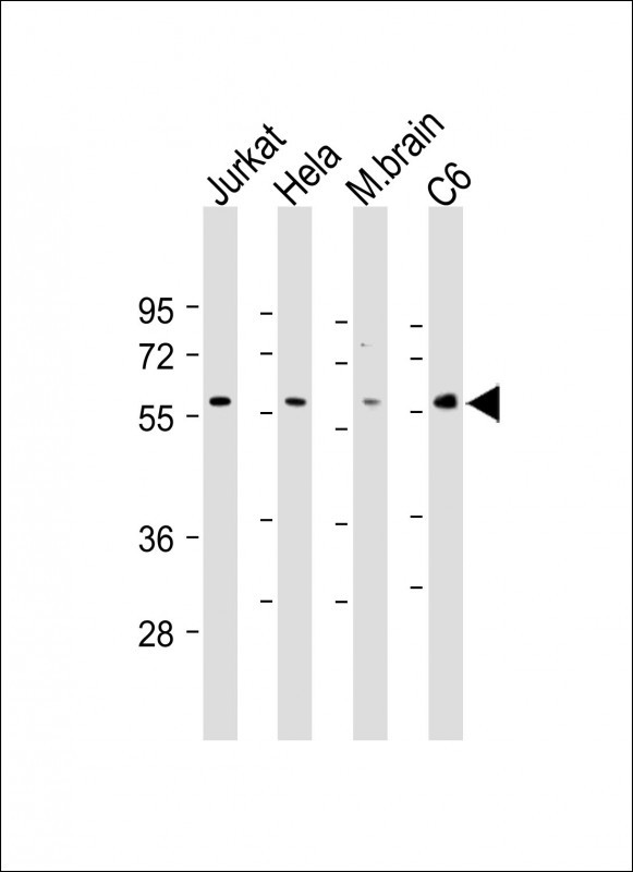
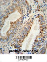
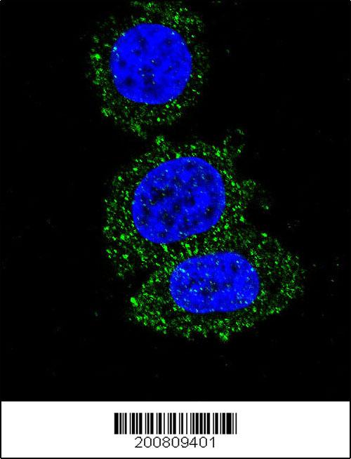
AKT2 Antibody
Purified Mouse Monoclonal Antibody (Mab)
- 产品详情
- 文献引用 : 2
- 实验流程
- 背景知识
Application
| WB, IHC-P, IF, E |
|---|---|
| Primary Accession | P31751 |
| Other Accession | NP_001617.1 |
| Reactivity | Human, Rat, Mouse |
| Host | Mouse |
| Clonality | Monoclonal |
| Isotype | Mouse IgG1 |
| Clone Names | 148CT5.4.1.4 |
| Calculated MW | 55769 Da |
| Gene ID | 208 |
|---|---|
| Other Names | RAC-beta serine/threonine-protein kinase, Protein kinase Akt-2, Protein kinase B beta, PKB beta, RAC-PK-beta, AKT2 |
| Target/Specificity | This AKT2 monoclonal antibody is generated from mouse immunized with AKT2 recombinant protein. |
| Dilution | WB~~1:500-1:2000 IHC-P~~1:100~500 IF~~1:10~50 E~~Use at an assay dependent concentration. |
| Format | Purified monoclonal antibody supplied in PBS with 0.09% (W/V) sodium azide. This antibody is purified through a protein G column, followed by dialysis against PBS. |
| Storage | Maintain refrigerated at 2-8°C for up to 2 weeks. For long term storage store at -20°C in small aliquots to prevent freeze-thaw cycles. |
| Precautions | AKT2 Antibody is for research use only and not for use in diagnostic or therapeutic procedures. |
| Name | AKT2 (HGNC:392) |
|---|---|
| Function | Serine/threonine kinase closely related to AKT1 and AKT3. All 3 enzymes, AKT1, AKT2 and AKT3, are collectively known as AKT kinase. AKT regulates many processes including metabolism, proliferation, cell survival, growth and angiogenesis, through the phosphorylation of a range of downstream substrates. Over 100 substrates have been reported so far, although for most of them, the precise AKT kinase catalyzing the reaction was not specified. AKT regulates glucose uptake by mediating insulin-induced translocation of the SLC2A4/GLUT4 glucose transporter to the cell surface. Phosphorylation of PTPN1 at 'Ser-50' negatively modulates its phosphatase activity preventing dephosphorylation of the insulin receptor and the attenuation of insulin signaling. Phosphorylation of TBC1D4 triggers the binding of this effector to inhibitory 14-3-3 proteins, which is required for insulin-stimulated glucose transport. AKT also regulates the storage of glucose in the form of glycogen by phosphorylating GSK3A at 'Ser-21' and GSK3B at 'Ser-9', resulting in inhibition of its kinase activity. Phosphorylation of GSK3 isoforms by AKT is also thought to be one mechanism by which cell proliferation is driven. AKT also regulates cell survival via the phosphorylation of MAP3K5 (apoptosis signal- related kinase). Phosphorylation of 'Ser-83' decreases MAP3K5 kinase activity stimulated by oxidative stress and thereby prevents apoptosis. AKT mediates insulin-stimulated protein synthesis by phosphorylating TSC2 at 'Ser-939' and 'Thr-1462', thereby activating mTORC1 signaling and leading to both phosphorylation of 4E-BP1 and in activation of RPS6KB1. AKT is involved in the phosphorylation of members of the FOXO factors (Forkhead family of transcription factors), leading to binding of 14-3-3 proteins and cytoplasmic localization. In particular, FOXO1 is phosphorylated at 'Thr-24', 'Ser-256' and 'Ser-319'. FOXO3 and FOXO4 are phosphorylated on equivalent sites. AKT has an important role in the regulation of NF-kappa-B-dependent gene transcription and positively regulates the activity of CREB1 (cyclic AMP (cAMP)-response element binding protein). The phosphorylation of CREB1 induces the binding of accessory proteins that are necessary for the transcription of pro-survival genes such as BCL2 and MCL1. AKT phosphorylates 'Ser- 454' on ATP citrate lyase (ACLY), thereby potentially regulating ACLY activity and fatty acid synthesis. Activates the 3B isoform of cyclic nucleotide phosphodiesterase (PDE3B) via phosphorylation of 'Ser-273', resulting in reduced cyclic AMP levels and inhibition of lipolysis. Phosphorylates PIKFYVE on 'Ser-318', which results in increased PI(3)P- 5 activity. The Rho GTPase-activating protein DLC1 is another substrate and its phosphorylation is implicated in the regulation cell proliferation and cell growth. AKT plays a role as key modulator of the AKT-mTOR signaling pathway controlling the tempo of the process of newborn neurons integration during adult neurogenesis, including correct neuron positioning, dendritic development and synapse formation. Signals downstream of phosphatidylinositol 3-kinase (PI(3)K) to mediate the effects of various growth factors such as platelet- derived growth factor (PDGF), epidermal growth factor (EGF), insulin and insulin-like growth factor 1 (IGF1). AKT mediates the antiapoptotic effects of IGF1. Essential for the SPATA13-mediated regulation of cell migration and adhesion assembly and disassembly. May be involved in the regulation of the placental development (PubMed:21432781, PubMed:21620960). In response to lysophosphatidic acid stimulation, inhibits the ciliogenesis cascade. In this context, phosphorylates WDR44, hence stabilizing its interaction with Rab11 and preventing the formation of the ciliogenic Rab11-FIP3-RAB3IP complex. Also phosphorylates RAB3IP/Rabin8, thus may affect RAB3IP guanine nucleotide exchange factor (GEF) activity toward Rab8, which is important for cilia growth (PubMed:31204173). Phosphorylates PKP1, facilitating its interaction with YWHAG and translocation to the nucleus, ultimately resulting in a reduction in keratinocyte intercellular adhesion (By similarity). Phosphorylation of PKP1 increases PKP1 protein stability, translocation to the cytoplasm away from desmosome plaques and PKP1- driven cap-dependent translation (PubMed:23444369). |
| Cellular Location | Cytoplasm. Nucleus Cell membrane; Peripheral membrane protein. Early endosome {ECO:0000250|UniProtKB:Q60823}. Note=Through binding of the N-terminal PH domain to phosphatidylinositol (3,4,5)-trisphosphate (PtdIns(3,4,5)P3) or phosphatidylinositol (3,4)-bisphosphate (PtdIns(3,4)P2), recruited to the plasma membrane. Cell membrane recruitment is facilitated by interaction with CLIP3. Colocalizes with WDFY2 in early endosomes (By similarity). Localizes within both nucleus and cytoplasm in proliferative primary myoblasts and mostly within the nucleus of differentiated primary myoblasts (PubMed:17565718) {ECO:0000250|UniProtKB:Q60823, ECO:0000269|PubMed:17565718} |
| Tissue Location | Widely expressed. Expressed in myoblasts (PubMed:17565718). |
For Research Use Only. Not For Use In Diagnostic Procedures.

Provided below are standard protocols that you may find useful for product applications.
BACKGROUND
AKT2 is a putative oncogene encoding a protein belonging to a subfamily of serine/threonine kinases containing SH2-like (Src homology 2-like) domains. AKT2 was shown to be amplified and overexpressed in 2 of 8 ovarian carcinoma cell lines and 2 of 15 primary ovarian tumors. Overexpression contributes to the malignant phenotype of a subset of human ductal pancreatic cancers. The encoded protein is a general protein kinase capable of phophorylating several known proteins.
REFERENCES
Fortier, A.M., et al. FEBS Lett. 584(5):984-988(2010)
Li, X., et al. Arterioscler. Thromb. Vasc. Biol. 30(3):582-590(2010)
Santi, S.A., et al. Am. J. Physiol., Cell Physiol. 298 (3), C580-C591 (2010)
终于等到您。ABCEPTA(百远生物)抗体产品。
点击下方“我要评价 ”按钮提交您的反馈信息,您的反馈和评价是我们最宝贵的财富之一,
我们将在1-3个工作日内处理您的反馈信息。
如有疑问,联系:0512-88856768 tech-china@abcepta.com.


