


 癌症的基本特征包括细胞增殖、血管生成、迁移、凋亡逃避机制和细胞永生等。找到癌症发生过程中这些通路的关键标记物和对应的抗体用于检测至关重要。
 为您推荐一个泛素化位点预测神器——泛素化分析工具,可以为您的蛋白的泛素化位点作出预测和评分。
 细胞自噬受体图形绘图工具为你的蛋白的细胞受体结合位点作出预测和评分,识别结合到自噬通路中的蛋白是非常重要的,便于让我们理解自噬在正常生理、病理过程中的作用,如发育、细胞分化、神经退化性疾病、压力条件下、感染和癌症。
LGALS3 Antibody (C-term)
Mouse Monoclonal Antibody (Mab)
- 产品详情
- 实验流程
- 背景知识
Application
| WB, IHC-P, E |
|---|---|
| Primary Accession | P17931 |
| Other Accession | NP_002297.2 |
| Reactivity | Human |
| Host | Mouse |
| Clonality | Monoclonal |
| Isotype | IgG1,k |
| Clone Names | 320CT4.5.2 |
| Calculated MW | 26152 Da |
| Antigen Region | 163-191 aa |
| Gene ID | 3958 |
|---|---|
| Other Names | Galectin-3, Gal-3, 35 kDa lectin, Carbohydrate-binding protein 35, CBP 35, Galactose-specific lectin 3, Galactoside-binding protein, GALBP, IgE-binding protein, L-31, Laminin-binding protein, Lectin L-29, Mac-2 antigen, LGALS3, MAC2 |
| Target/Specificity | This LGALS3 antibody is generated from mice immunized with a KLH conjugated synthetic peptide between 163-191 amino acids from the C-terminal region of human LGALS3. |
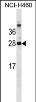
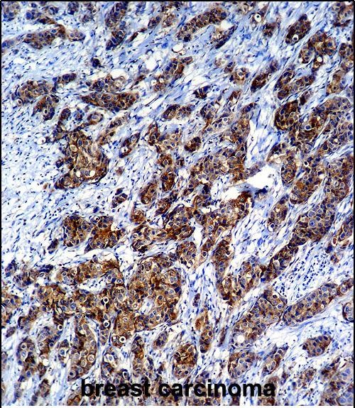
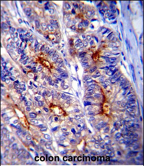
| Dilution | WB~~1:500~1000 IHC-P~~1:100~500 E~~Use at an assay dependent concentration. |
| Format | Purified monoclonal antibody supplied in PBS with 0.09% (W/V) sodium azide. This antibody is purified through a protein G column, followed by dialysis against PBS. |
| Storage | Maintain refrigerated at 2-8°C for up to 2 weeks. For long term storage store at -20°C in small aliquots to prevent freeze-thaw cycles. |
| Precautions | LGALS3 Antibody (C-term) is for research use only and not for use in diagnostic or therapeutic procedures. |
| Name | LGALS3 (HGNC:6563) |
|---|---|
| Synonyms | MAC2 |
| Function | Galactose-specific lectin which binds IgE. May mediate with the alpha-3, beta-1 integrin the stimulation by CSPG4 of endothelial cells migration. Together with DMBT1, required for terminal differentiation of columnar epithelial cells during early embryogenesis (By similarity). In the nucleus: acts as a pre-mRNA splicing factor. Involved in acute inflammatory responses including neutrophil activation and adhesion, chemoattraction of monocytes macrophages, opsonization of apoptotic neutrophils, and activation of mast cells. Together with TRIM16, coordinates the recognition of membrane damage with mobilization of the core autophagy regulators ATG16L1 and BECN1 in response to damaged endomembranes. |
| Cellular Location | Cytoplasm. Nucleus. Secreted. Note=Secreted by a non- classical secretory pathway and associates with the cell surface. Can be secreted; the secretion is dependent on protein unfolding and facilitated by the cargo receptor TMED10; it results in protein translocation from the cytoplasm into the ERGIC (endoplasmic reticulum- Golgi intermediate compartment) followed by vesicle entry and secretion (PubMed:32272059). |
| Tissue Location | A major expression is found in the colonic epithelium. It is also abundant in the activated macrophages. Expressed in fetal membranes. |
For Research Use Only. Not For Use In Diagnostic Procedures.
Provided below are standard protocols that you may find useful for product applications.
BACKGROUND
This gene encodes a member of the galectin family of carbohydrate binding proteins. Members of this protein family have an affinity for beta-galactosides. The encoded protein is characterized by an N-terminal proline-rich tandem repeat domain and a single C-terminal carbohydrate recognition domain. This protein can self-associate through the N-terminal domain allowing it to bind to multivalent saccharide ligands. This protein localizes to the extracellular matrix, the cytoplasm and the nucleus. This protein plays a role in numerous cellular functions including apoptosis, innate immunity, cell adhesion and T-cell regulation. Alternate splicing results in multiple transcript variants.
REFERENCES
Salomonsson, E., et al. J. Biol. Chem. 285(45):35079-35091(2010)
Debierre-Grockiego, F., et al. J. Biol. Chem. 285(43):32744-32750(2010)
Zhou, J.Y., et al. J. Proteome Res. 9(10):5133-5141(2010)
Markowska, A.I., et al. J. Exp. Med. 207(9):1981-1993(2010)
Mazurek, N., et al. J. Biol. Chem. 275(46):36311-36315(2000)
终于等到您。ABCEPTA(百远生物)抗体产品。
点击下方“我要评价 ”按钮提交您的反馈信息,您的反馈和评价是我们最宝贵的财富之一,
我们将在1-3个工作日内处理您的反馈信息。
如有疑问,联系:0512-88856768 tech-china@abcepta.com.


