


 癌症的基本特征包括细胞增殖、血管生成、迁移、凋亡逃避机制和细胞永生等。找到癌症发生过程中这些通路的关键标记物和对应的抗体用于检测至关重要。
 为您推荐一个泛素化位点预测神器——泛素化分析工具,可以为您的蛋白的泛素化位点作出预测和评分。
 细胞自噬受体图形绘图工具为你的蛋白的细胞受体结合位点作出预测和评分,识别结合到自噬通路中的蛋白是非常重要的,便于让我们理解自噬在正常生理、病理过程中的作用,如发育、细胞分化、神经退化性疾病、压力条件下、感染和癌症。
PSMA5 Antibody
Mouse Monoclonal Antibody (Mab)
- 产品详情
- 实验流程
- 背景知识
Application
| WB, IHC-P, IF, E |
|---|---|
| Primary Accession | P28066 |
| Other Accession | Q9Z2U1, Q5E987, NP_002781.2 |
| Reactivity | Human, Rat, Mouse |
| Predicted | Bovine, Mouse |
| Host | Mouse |
| Clonality | Monoclonal |
| Isotype | IgG1 |
| Clone Names | 426CT8.5.1 |
| Calculated MW | 26411 Da |
| Gene ID | 5686 |
|---|---|
| Other Names | Proteasome subunit alpha type-5, Macropain zeta chain, Multicatalytic endopeptidase complex zeta chain, Proteasome zeta chain, PSMA5 |
| Target/Specificity | Purified His-tagged PSMA5 protein(Fragment) was used to produced this monoclonal antibody. |
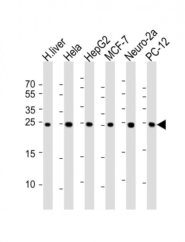
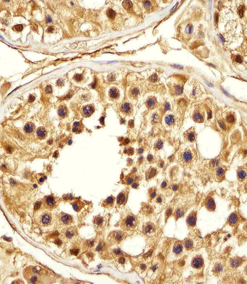
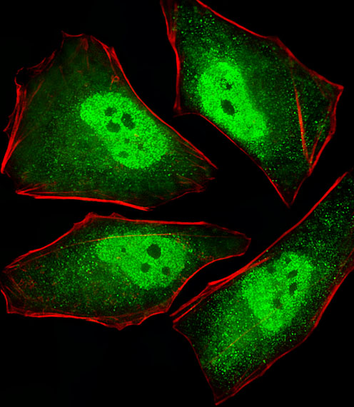
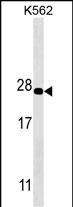
| Dilution | WB~~1:500~1000 IHC-P~~1:100~500 IF~~1:25 E~~Use at an assay dependent concentration. |
| Format | Purified monoclonal antibody supplied in PBS with 0.09% (W/V) sodium azide. This antibody is purified through a protein G column, followed by dialysis against PBS. |
| Storage | Maintain refrigerated at 2-8°C for up to 2 weeks. For long term storage store at -20°C in small aliquots to prevent freeze-thaw cycles. |
| Precautions | PSMA5 Antibody is for research use only and not for use in diagnostic or therapeutic procedures. |
| Name | PSMA5 (HGNC:9534) |
|---|---|
| Function | Component of the 20S core proteasome complex involved in the proteolytic degradation of most intracellular proteins. This complex plays numerous essential roles within the cell by associating with different regulatory particles. Associated with two 19S regulatory particles, forms the 26S proteasome and thus participates in the ATP- dependent degradation of ubiquitinated proteins. The 26S proteasome plays a key role in the maintenance of protein homeostasis by removing misfolded or damaged proteins that could impair cellular functions, and by removing proteins whose functions are no longer required. Associated with the PA200 or PA28, the 20S proteasome mediates ubiquitin- independent protein degradation. This type of proteolysis is required in several pathways including spermatogenesis (20S-PA200 complex) or generation of a subset of MHC class I-presented antigenic peptides (20S-PA28 complex). |
| Cellular Location | Cytoplasm. Nucleus. Note=Translocated from the cytoplasm into the nucleus following interaction with AKIRIN2, which bridges the proteasome with the nuclear import receptor IPO9 |
| Tissue Location | Expressed in fetal brain (at protein level). |
For Research Use Only. Not For Use In Diagnostic Procedures.
Provided below are standard protocols that you may find useful for product applications.
BACKGROUND
The proteasome is a multicatalytic proteinase complex with a highly ordered ring-shaped 20S core structure. The core structure is composed of 4 rings of 28 non-identical subunits; 2 rings are composed of 7 alpha subunits and 2 rings are composed of 7 beta subunits. Proteasomes are distributed throughout eukaryotic cells at a high concentration and cleave peptides in an ATP/ubiquitin-dependent process in a non-lysosomal pathway. An essential function of a modified proteasome, the immunoproteasome, is the processing of class I MHC peptides. This gene encodes a member of the peptidase T1A family, that is a 20S core alpha subunit.
REFERENCES
Kottgen, A., et al. Nat. Genet. 42(5):376-384(2010)
Sugiyama, N., et al. Mol. Cell Proteomics 6(6):1103-1109(2007)
Olsen, J.V., et al. Cell 127(3):635-648(2006)
Beausoleil, S.A., et al. Nat. Biotechnol. 24(10):1285-1292(2006)
Hirano, Y., et al. Nature 437(7063):1381-1385(2005)
终于等到您。ABCEPTA(百远生物)抗体产品。
点击下方“我要评价 ”按钮提交您的反馈信息,您的反馈和评价是我们最宝贵的财富之一,
我们将在1-3个工作日内处理您的反馈信息。
如有疑问,联系:0512-88856768 tech-china@abcepta.com.


