


 癌症的基本特征包括细胞增殖、血管生成、迁移、凋亡逃避机制和细胞永生等。找到癌症发生过程中这些通路的关键标记物和对应的抗体用于检测至关重要。
 为您推荐一个泛素化位点预测神器——泛素化分析工具,可以为您的蛋白的泛素化位点作出预测和评分。
 细胞自噬受体图形绘图工具为你的蛋白的细胞受体结合位点作出预测和评分,识别结合到自噬通路中的蛋白是非常重要的,便于让我们理解自噬在正常生理、病理过程中的作用,如发育、细胞分化、神经退化性疾病、压力条件下、感染和癌症。
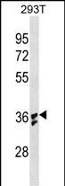
IL1B Antibody (Center) (Ascites)
Mouse Monoclonal Antibody (Mab)
- 产品详情
- 文献引用 : 1
- 实验流程
- 背景知识
Application
| WB, E |
|---|---|
| Primary Accession | P01584 |
| Other Accession | P14628, P79182, NP_000567.1 |
| Reactivity | Human |
| Predicted | Monkey, Rabbit |
| Host | Mouse |
| Clonality | Monoclonal |
| Isotype | IgG1 |
| Clone Names | 614CT4.3.1 |
| Calculated MW | 30748 Da |
| Antigen Region | 148-174 aa |
| Gene ID | 3553 |
|---|---|
| Other Names | Interleukin-1 beta, IL-1 beta, Catabolin, IL1B, IL1F2 |
| Target/Specificity | This IL1B antibody is generated from mice immunized with a KLH conjugated synthetic peptide between 148-174 amino acids from the Central region of human IL1B. |
| Dilution | WB~~1:2000~4000 E~~Use at an assay dependent concentration. |
| Format | Mouse monoclonal antibody supplied in crude ascites with 0.09% (W/V) sodium azide. |
| Storage | Maintain refrigerated at 2-8°C for up to 2 weeks. For long term storage store at -20°C in small aliquots to prevent freeze-thaw cycles. |
| Precautions | IL1B Antibody (Center) (Ascites) is for research use only and not for use in diagnostic or therapeutic procedures. |
| Name | IL1B (HGNC:5992) |
|---|---|
| Synonyms | IL1F2 |
| Function | Potent pro-inflammatory cytokine (PubMed:10653850, PubMed:12794819, PubMed:28331908, PubMed:3920526). Initially discovered as the major endogenous pyrogen, induces prostaglandin synthesis, neutrophil influx and activation, T-cell activation and cytokine production, B-cell activation and antibody production, and fibroblast proliferation and collagen production (PubMed:3920526). Promotes Th17 differentiation of T-cells. Synergizes with IL12/interleukin-12 to induce IFNG synthesis from T-helper 1 (Th1) cells (PubMed:10653850). Plays a role in angiogenesis by inducing VEGF production synergistically with TNF and IL6 (PubMed:12794819). Involved in transduction of inflammation downstream of pyroptosis: its mature form is specifically released in the extracellular milieu by passing through the gasdermin-D (GSDMD) pore (PubMed:33377178, PubMed:33883744). Acts as a sensor of S.pyogenes infection in skin: cleaved and activated by pyogenes SpeB protease, leading to an inflammatory response that prevents bacterial growth during invasive skin infection (PubMed:28331908). |
| Cellular Location | Cytoplasm, cytosol. Secreted. Lysosome Secreted, extracellular exosome {ECO:0000250|UniProtKB:P10749} Note=The precursor is cytosolic (PubMed:15192144). In response to inflammasome-activating signals, such as ATP for NLRP3 inflammasome or bacterial flagellin for NLRC4 inflammasome, cleaved and secreted (PubMed:24201029, PubMed:33377178, PubMed:33883744). Mature form is secreted and released in the extracellular milieu by passing through the gasdermin-D (GSDMD) pore (PubMed:33883744). In contrast, the precursor form is not released, due to the presence of an acidic region that is proteolytically removed by CASP1 during maturation (PubMed:33883744). The secretion is dependent on protein unfolding and facilitated by the cargo receptor TMED10 (PubMed:32272059) |
| Tissue Location | Expressed in activated monocytes/macrophages (at protein level). |
For Research Use Only. Not For Use In Diagnostic Procedures.

Provided below are standard protocols that you may find useful for product applications.
BACKGROUND
The protein encoded by this gene is a member of the interleukin 1 cytokine family. This cytokine is produced by activated macrophages as a proprotein, which is proteolytically processed to its active form by caspase 1 (CASP1/ICE). This cytokine is an important mediator of the inflammatory response, and is involved in a variety of cellular activities, including cell proliferation, differentiation, and apoptosis. The induction of cyclooxygenase-2 (PTGS2/COX2) by this cytokine in the central nervous system (CNS) is found to contribute to inflammatory pain hypersensitivity. This gene and eight other interleukin 1 family genes form a cytokine gene cluster on chromosome 2. [provided by RefSeq].
REFERENCES
Lee, B., et al. J. Immunol. 185(10):5926-5934(2010)
Arana-Argaez, V.E., et al. J. Biol. Chem. 285(43):32824-32833(2010)
Zhang, Z., et al. J. Biol. Chem. 285(43):33092-33103(2010)
Wang, D., et al. Nat. Immunol. 11(10):905-911(2010)
Gein, O.N., et al. Patol Fiziol Eksp Ter 1, 10-13 (2010) :
终于等到您。ABCEPTA(百远生物)抗体产品。
点击下方“我要评价 ”按钮提交您的反馈信息,您的反馈和评价是我们最宝贵的财富之一,
我们将在1-3个工作日内处理您的反馈信息。
如有疑问,联系:0512-88856768 tech-china@abcepta.com.


