


 癌症的基本特征包括细胞增殖、血管生成、迁移、凋亡逃避机制和细胞永生等。找到癌症发生过程中这些通路的关键标记物和对应的抗体用于检测至关重要。
 为您推荐一个泛素化位点预测神器——泛素化分析工具,可以为您的蛋白的泛素化位点作出预测和评分。
 细胞自噬受体图形绘图工具为你的蛋白的细胞受体结合位点作出预测和评分,识别结合到自噬通路中的蛋白是非常重要的,便于让我们理解自噬在正常生理、病理过程中的作用,如发育、细胞分化、神经退化性疾病、压力条件下、感染和癌症。
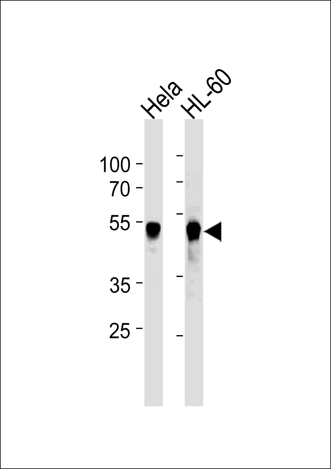
VRK1 Antibody (Center)(Ascites)
Mouse Monoclonal Antibody (Mab)
- 产品详情
- 实验流程
- 背景知识
Application
| WB, E |
|---|---|
| Primary Accession | Q99986 |
| Reactivity | Human |
| Host | Mouse |
| Clonality | Monoclonal |
| Isotype | IgG1 |
| Clone Names | 1015CT2.1.1 |
| Calculated MW | 45476 Da |
| Gene ID | 7443 |
|---|---|
| Other Names | Serine/threonine-protein kinase VRK1, Vaccinia-related kinase 1, VRK1 |
| Target/Specificity | Purified His-tagged VRK1 protein was used to produced this monoclonal antibody. |
| Dilution | WB~~1:5000 E~~Use at an assay dependent concentration. |
| Format | Mouse monoclonal antibody supplied in crude ascites with 0.09% (W/V) sodium azide. |
| Storage | Maintain refrigerated at 2-8°C for up to 2 weeks. For long term storage store at -20°C in small aliquots to prevent freeze-thaw cycles. |
| Precautions | VRK1 Antibody (Center)(Ascites) is for research use only and not for use in diagnostic or therapeutic procedures. |
| Name | VRK1 {ECO:0000303|PubMed:9344656, ECO:0000312|HGNC:HGNC:12718} |
|---|---|
| Function | Serine/threonine kinase involved in the regulation of key cellular processes including the cell cycle, nuclear condensation, transcription regulation, and DNA damage response (PubMed:14645249, PubMed:18617507, PubMed:19103756, PubMed:33076429). Controls chromatin organization and remodeling by mediating phosphorylation of histone H3 on 'Thr-4' and histone H2AX (H2aXT4ph) (PubMed:31527692, PubMed:37179361). It also phosphorylates KAT5 in response to DNA damage, promoting KAT5 association with chromatin and histone acetyltransferase activity (PubMed:33076429). Is involved in the regulation of cell cycle progression of neural progenitors, and is required for proper cortical neuronal migration (By similarity). Is involved in neurite elongation and branching in motor neurons, and has an essential role in Cajal bodies assembly, acting through COIL phosphorylation and the control of coilin degradation (PubMed:21920476, PubMed:31090908, PubMed:31527692). Involved in Golgi disassembly during the cell cycle: following phosphorylation by PLK3 during mitosis, it is required to induce Golgi fragmentation (PubMed:19103756). Phosphorylates BANF1: disrupts its ability to bind DNA, reduces its binding to LEM domain-containing proteins and causes its relocalization from the nucleus to the cytoplasm (PubMed:16495336). Phosphorylates TP53BP1 and p53/TP53 on 'Thr-18', preventing the interaction between p53/TP53 and MDM2 (PubMed:10951572, PubMed:31527692). Phosphorylates ATF2 which activates its transcriptional activity (PubMed:15105425). Phosphorylates JUN (PubMed:31527692). |
| Cellular Location | Nucleus. Cytoplasm. Nucleus, Cajal body. Note=Enriched on chromatin during mitosis. |
| Tissue Location | Widely expressed. Highly expressed in fetal liver, testis and thymus. |
For Research Use Only. Not For Use In Diagnostic Procedures.
Provided below are standard protocols that you may find useful for product applications.
BACKGROUND
Serine/threonine kinase involved in Golgi disassembly during the cell cycle: following phosphorylation by PLK3 during mitosis, required to induce Golgi fragmentation. Acts by mediating phosphorylation of downstream target protein. Phosphorylates 'Thr-18' of p53/TP53 and may thereby prevent the interaction between p53/TP53 and MDM2. Phosphorylates casein and histone H3. Phosphorylates BANF1: disrupts its ability to bind DNA, reduces its binding to LEM domain-containing proteins and causes its relocalization from the nucleus to the cytoplasm.
REFERENCES
Nezu J., et al. Genomics 45:327-331(1997).
Lopez-Borges S., et al. Oncogene 19:3656-3664(2000).
Barcia R., et al. Arch. Biochem. Biophys. 399:1-5(2002).
Nichols R.J., et al. J. Biol. Chem. 279:7934-7946(2004).
Blanco S., et al. FEBS J. 273:2487-2504(2006).
终于等到您。ABCEPTA(百远生物)抗体产品。
点击下方“我要评价 ”按钮提交您的反馈信息,您的反馈和评价是我们最宝贵的财富之一,
我们将在1-3个工作日内处理您的反馈信息。
如有疑问,联系:0512-88856768 tech-china@abcepta.com.


