


 癌症的基本特征包括细胞增殖、血管生成、迁移、凋亡逃避机制和细胞永生等。找到癌症发生过程中这些通路的关键标记物和对应的抗体用于检测至关重要。
 为您推荐一个泛素化位点预测神器——泛素化分析工具,可以为您的蛋白的泛素化位点作出预测和评分。
 细胞自噬受体图形绘图工具为你的蛋白的细胞受体结合位点作出预测和评分,识别结合到自噬通路中的蛋白是非常重要的,便于让我们理解自噬在正常生理、病理过程中的作用,如发育、细胞分化、神经退化性疾病、压力条件下、感染和癌症。
RAC1 Antibody
Purified Mouse Monoclonal Antibody (Mab)
- 产品详情
- 实验流程
- 背景知识
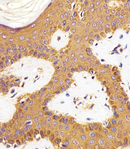
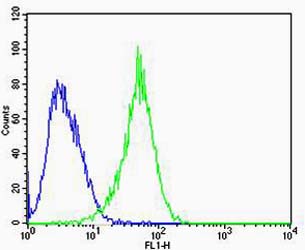
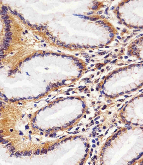
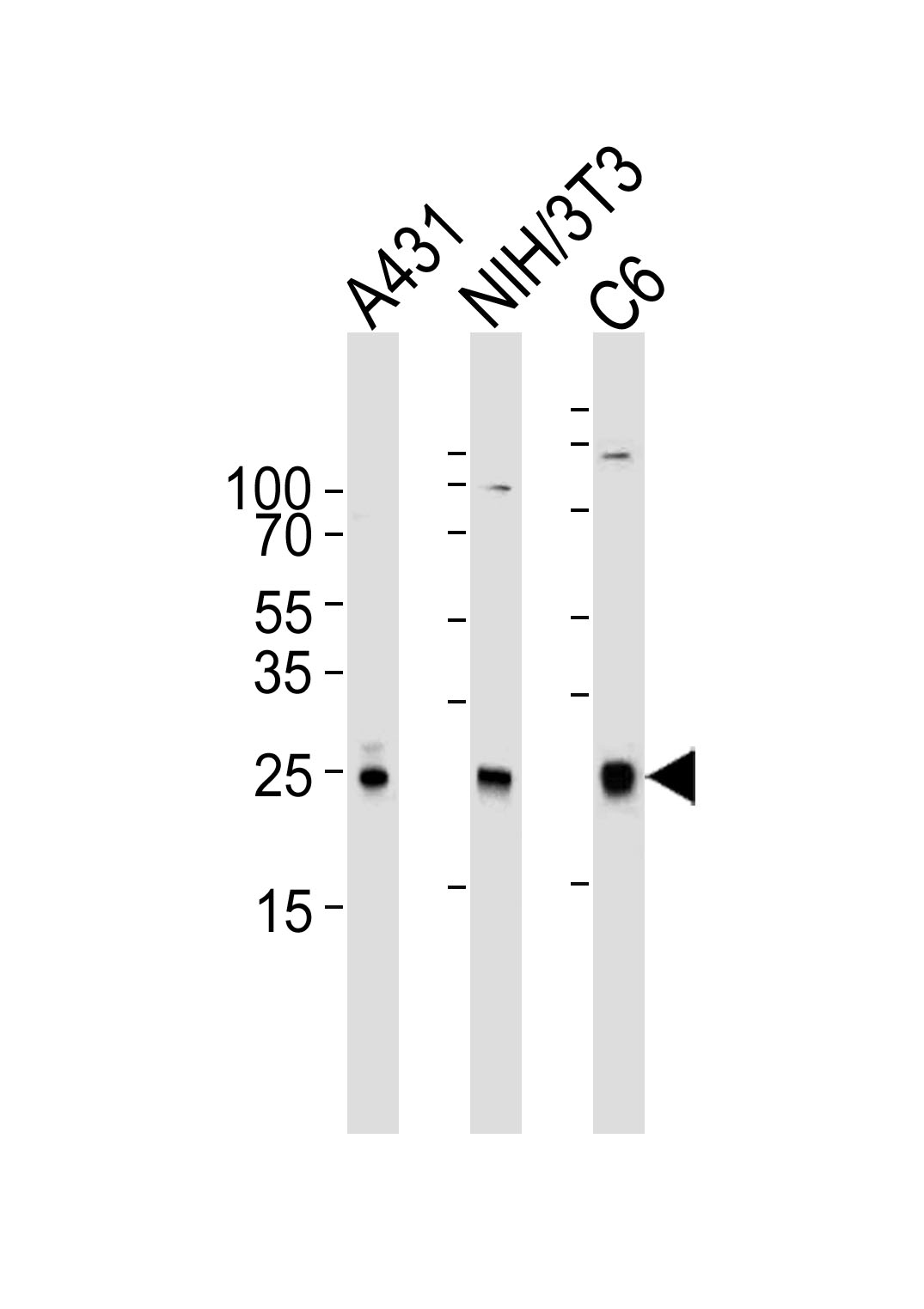
Application
| WB, IHC-P, FC, E |
|---|---|
| Primary Accession | P63000 |
| Reactivity | Human, Rat, Mouse |
| Host | Mouse |
| Clonality | Monoclonal |
| Isotype | IgG2b |
| Clone Names | 1301CT276.121.104 |
| Calculated MW | 21450 Da |
| Gene ID | 5879 |
|---|---|
| Other Names | Ras-related C3 botulinum toxin substrate 1, Cell migration-inducing gene 5 protein, Ras-like protein TC25, p21-Rac1, RAC1, TC25 |
| Target/Specificity | This antibody is generated from a mouse immunized with a KLH conjugated synthetic peptide between amino acids from human. |
| Dilution | WB~~1:1000 IHC-P~~1:100~500 FC~~1:100 E~~Use at an assay dependent concentration. |
| Format | Purified monoclonal antibody supplied in PBS with 0.09% (W/V) sodium azide. This antibody is purified through a protein G column, followed by dialysis against PBS. |
| Storage | Maintain refrigerated at 2-8°C for up to 2 weeks. For long term storage store at -20°C in small aliquots to prevent freeze-thaw cycles. |
| Precautions | RAC1 Antibody is for research use only and not for use in diagnostic or therapeutic procedures. |
| Name | RAC1 (HGNC:9801) |
|---|---|
| Synonyms | TC25 |
| Function | Plasma membrane-associated small GTPase which cycles between active GTP-bound and inactive GDP-bound states. In its active state, binds to a variety of effector proteins to regulate cellular responses such as secretory processes, phagocytosis of apoptotic cells, epithelial cell polarization, neurons adhesion, migration and differentiation, and growth-factor induced formation of membrane ruffles (PubMed:1643658, PubMed:22843693, PubMed:23512198, PubMed:28886345). Rac1 p21/rho GDI heterodimer is the active component of the cytosolic factor sigma 1, which is involved in stimulation of the NADPH oxidase activity in macrophages. Essential for the SPATA13- mediated regulation of cell migration and adhesion assembly and disassembly. Stimulates PKN2 kinase activity (PubMed:9121475). In concert with RAB7A, plays a role in regulating the formation of RBs (ruffled borders) in osteoclasts (PubMed:1643658). In podocytes, promotes nuclear shuttling of NR3C2; this modulation is required for a proper kidney functioning. Required for atypical chemokine receptor ACKR2-induced LIMK1-PAK1-dependent phosphorylation of cofilin (CFL1) and for up-regulation of ACKR2 from endosomal compartment to cell membrane, increasing its efficiency in chemokine uptake and degradation. In neurons, is involved in dendritic spine formation and synaptic plasticity (By similarity). In hippocampal neurons, involved in spine morphogenesis and synapse formation, through local activation at synapses by guanine nucleotide exchange factors (GEFs), such as ARHGEF6/ARHGEF7/PIX (PubMed:12695502). In synapses, seems to mediate the regulation of F-actin cluster formation performed by SHANK3. In neurons, plays a crucial role in regulating GABA(A) receptor synaptic stability and hence GABAergic inhibitory synaptic transmission through its role in PAK1 activation and eventually F-actin stabilization (By similarity). Required for DSG3 translocation to cell-cell junctions, DSG3-mediated organization of cortical F-actin bundles and anchoring of actin at cell junctions; via interaction with DSG3 (PubMed:22796473). Subunit of the phagocyte NADPH oxidase complex that mediates the transfer of electrons from cytosolic NADPH to O2 to produce the superoxide anion (O2(-)) (PubMed:38355798). |
| Cellular Location | Cell membrane; Lipid-anchor; Cytoplasmic side. Melanosome. Cytoplasm. Cell projection, lamellipodium {ECO:0000250|UniProtKB:P63001}. Cell projection, dendrite {ECO:0000250|UniProtKB:P63001}. Synapse {ECO:0000250|UniProtKB:Q6RUV5} Nucleus. Note=Inner surface of plasma membrane possibly with attachment requiring prenylation of the C- terminal cysteine (PubMed:1903399). Identified by mass spectrometry in melanosome fractions from stage I to stage IV (PubMed:17081065). Found in the ruffled border (a late endosomal-like compartment in the plasma membrane) of bone-resorbing osteoclasts. Localizes to the lamellipodium in a SH3RF1-dependent manner (By similarity). In macrophages, cytoplasmic location increases upon CSF1 stimulation (By similarity) Activation by GTP-binding promotes nuclear localization (PubMed:12551911). {ECO:0000250|UniProtKB:P63001, ECO:0000250|UniProtKB:Q6RUV5, ECO:0000269|PubMed:12551911, ECO:0000269|PubMed:17081065, ECO:0000269|PubMed:1903399} |
| Tissue Location | Isoform B is predominantly identified in skin and epithelial tissues from the intestinal tract. Its expression is elevated in colorectal tumors at various stages of neoplastic progression, as compared to their respective adjacent tissues |
For Research Use Only. Not For Use In Diagnostic Procedures.
Provided below are standard protocols that you may find useful for product applications.
BACKGROUND
Plasma membrane-associated small GTPase which cycles between active GTP-bound and inactive GDP-bound states. In its active state, binds to a variety of effector proteins to regulate cellular responses such as secretory processes, phagocytosis of apoptotic cells, epithelial cell polarization and growth-factor induced formation of membrane ruffles. Rac1 p21/rho GDI heterodimer is the active component of the cytosolic factor sigma 1, which is involved in stimulation of the NADPH oxidase activity in macrophages. Essential for the SPATA13-mediated regulation of cell migration and adhesion assembly and disassembly. Stimulates PKN2 kinase activity. In concert with RAB7A, plays a role in regulating the formation of RBs (ruffled borders) in osteoclasts. In glioma cells, promotes cell migration and invasion. In podocytes, promotes nuclear shuttling of NR3C2; this modulation is required for a proper kidney functioning. Required for atypical chemokine receptor ACKR2-induced LIMK1-PAK1-dependent phosphorylation of cofilin (CFL1) and for up-regulation of ACKR2 from endosomal compartment to cell membrane, increasing its efficiency in chemokine uptake and degradation. In synapses, seems to mediate the regulation of F-actin cluster formation performed by SHANK3.
REFERENCES
Didsbury J.,et al.J. Biol. Chem. 264:16378-16382(1989).
Drivas G.T.,et al.Mol. Cell. Biol. 10:1793-1798(1990).
Matos P.,et al.Biochem. Biophys. Res. Commun. 277:741-751(2000).
Jordan P.,et al.Oncogene 18:6835-6839(1999).
Schnelzer A.,et al.Submitted (MAR-1999) to the EMBL/GenBank/DDBJ databases.
终于等到您。ABCEPTA(百远生物)抗体产品。
点击下方“我要评价 ”按钮提交您的反馈信息,您的反馈和评价是我们最宝贵的财富之一,
我们将在1-3个工作日内处理您的反馈信息。
如有疑问,联系:0512-88856768 tech-china@abcepta.com.


