


 癌症的基本特征包括细胞增殖、血管生成、迁移、凋亡逃避机制和细胞永生等。找到癌症发生过程中这些通路的关键标记物和对应的抗体用于检测至关重要。
 为您推荐一个泛素化位点预测神器——泛素化分析工具,可以为您的蛋白的泛素化位点作出预测和评分。
 细胞自噬受体图形绘图工具为你的蛋白的细胞受体结合位点作出预测和评分,识别结合到自噬通路中的蛋白是非常重要的,便于让我们理解自噬在正常生理、病理过程中的作用,如发育、细胞分化、神经退化性疾病、压力条件下、感染和癌症。
IRF3 Antibody
Purified Mouse Monoclonal Antibody (Mab)
- 产品详情
- 实验流程
- 背景知识
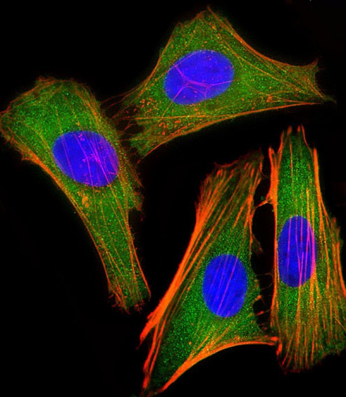
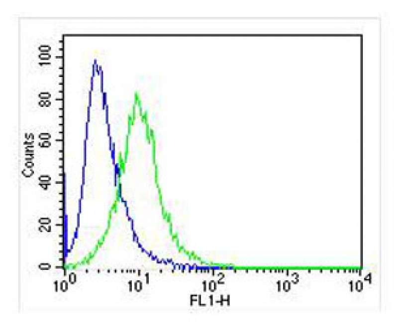
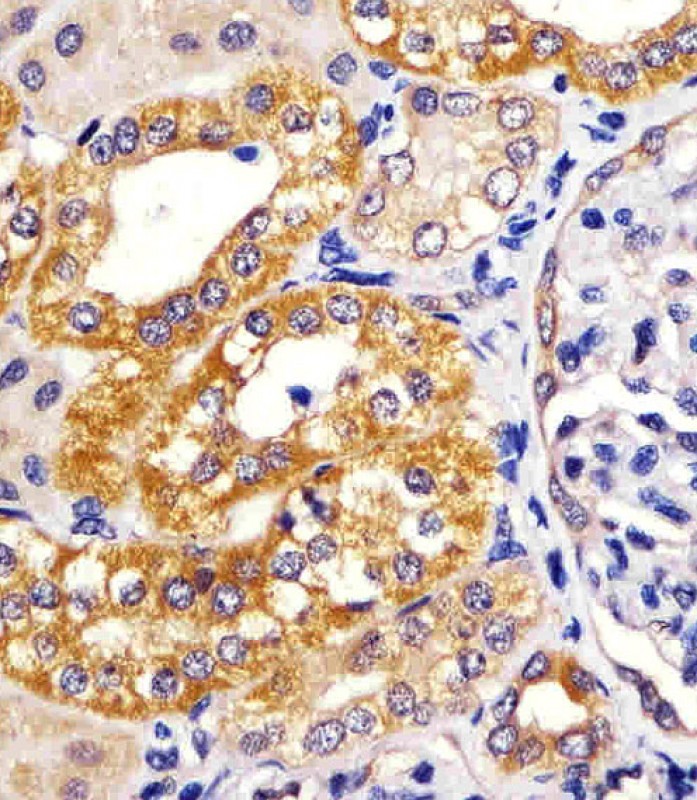
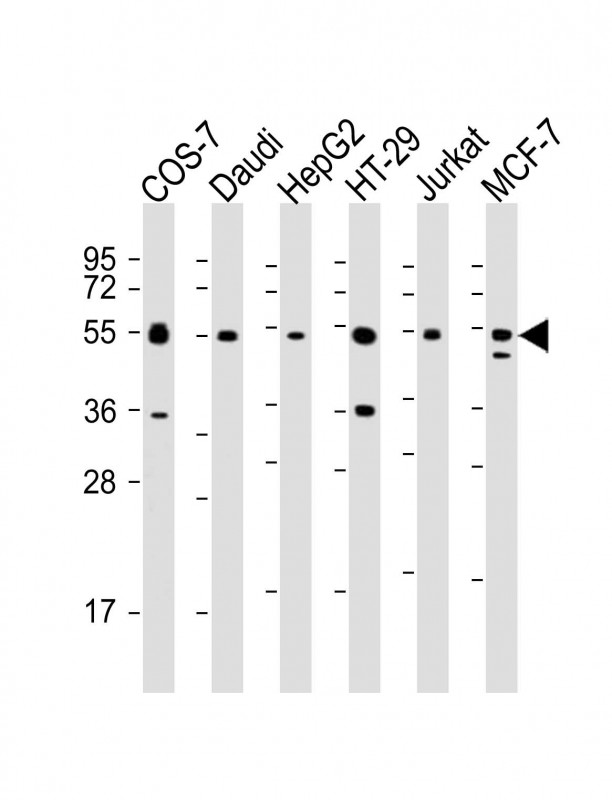
Application
| WB, IHC-P, FC, IF, E |
|---|---|
| Primary Accession | Q14653 |
| Reactivity | Human, Green Monkey, Mouse |
| Host | Mouse |
| Clonality | monoclonal |
| Isotype | IgG1,k |
| Clone Names | 1522CT766.58.24 |
| Calculated MW | 47219 Da |
| Gene ID | 3661 |
|---|---|
| Other Names | Interferon regulatory factor 3, IRF-3, IRF3 |
| Target/Specificity | This IRF3 antibody is generated from a mouse immunized with a recombinant protein. |
| Dilution | WB~~1:2000 IHC-P~~1:100~500 FC~~1:25 IF~~1:25 E~~Use at an assay dependent concentration. |
| Format | Purified monoclonal antibody supplied in PBS with 0.09% (W/V) sodium azide. This antibody is purified through a protein G column, followed by dialysis against PBS. |
| Storage | Maintain refrigerated at 2-8°C for up to 2 weeks. For long term storage store at -20°C in small aliquots to prevent freeze-thaw cycles. |
| Precautions | IRF3 Antibody is for research use only and not for use in diagnostic or therapeutic procedures. |
| Name | IRF3 {ECO:0000303|PubMed:9803267, ECO:0000312|HGNC:HGNC:6118} |
|---|---|
| Function | Key transcriptional regulator of type I interferon (IFN)- dependent immune responses which plays a critical role in the innate immune response against DNA and RNA viruses (PubMed:22394562, PubMed:24049179, PubMed:25636800, PubMed:27302953, PubMed:31340999, PubMed:36603579, PubMed:8524823, PubMed:39362857). Regulates the transcription of type I IFN genes (IFN-alpha and IFN-beta) and IFN- stimulated genes (ISG) by binding to an interferon-stimulated response element (ISRE) in their promoters (PubMed:11846977, PubMed:16846591, PubMed:16979567, PubMed:20049431, PubMed:32972995, PubMed:36603579, PubMed:8524823). Acts as a more potent activator of the IFN-beta (IFNB) gene than the IFN-alpha (IFNA) gene and plays a critical role in both the early and late phases of the IFNA/B gene induction (PubMed:16846591, PubMed:16979567, PubMed:20049431, PubMed:36603579). Found in an inactive form in the cytoplasm of uninfected cells and following viral infection, double-stranded RNA (dsRNA), or toll-like receptor (TLR) signaling, is phosphorylated by IKBKE and TBK1 kinases (PubMed:22394562, PubMed:25636800, PubMed:27302953, PubMed:36603579). This induces a conformational change, leading to its dimerization and nuclear localization and association with CREB binding protein (CREBBP) to form dsRNA-activated factor 1 (DRAF1), a complex which activates the transcription of the type I IFN and ISG genes (PubMed:16154084, PubMed:27302953, PubMed:33440148, PubMed:36603579). Can activate distinct gene expression programs in macrophages and can induce significant apoptosis in primary macrophages (PubMed:16846591). In response to Sendai virus infection, is recruited by TOMM70:HSP90AA1 to mitochondrion and forms an apoptosis complex TOMM70:HSP90AA1:IRF3:BAX inducing apoptosis (PubMed:25609812). Key transcription factor regulating the IFN response during SARS-CoV-2 infection (PubMed:33440148). |
| Cellular Location | Cytoplasm. Nucleus Mitochondrion. Note=Shuttles between cytoplasmic and nuclear compartments, with export being the prevailing effect (PubMed:10805757, PubMed:35922005). When activated, IRF3 interaction with CREBBP prevents its export to the cytoplasm (PubMed:10805757) Recruited to mitochondria via TOMM70:HSP90AA1 upon Sendai virus infection (PubMed:25609812). |
| Tissue Location | Expressed constitutively in a variety of tissues. |
For Research Use Only. Not For Use In Diagnostic Procedures.
Provided below are standard protocols that you may find useful for product applications.
BACKGROUND
Key transcriptional regulator of type I interferon (IFN)-dependent immune responses which plays a critical role in the innate immune response against DNA and RNA viruses. Regulates the transcription of type I IFN genes (IFN-alpha and IFN-beta) and IFN-stimulated genes (ISG) by binding to an interferon-stimulated response element (ISRE) in their promoters. Acts as a more potent activator of the IFN-beta (IFNB) gene than the IFN-alpha (IFNA) gene and plays a critical role in both the early and late phases of the IFNA/B gene induction. Found in an inactive form in the cytoplasm of uninfected cells and following viral infection, double-stranded RNA (dsRNA), or toll-like receptor (TLR) signaling, is phosphorylated by IKBKE and TBK1 kinases. This induces a conformational change, leading to its dimerization and nuclear localization and association with CREB binding protein (CREBBP) to form dsRNA-activated factor 1 (DRAF1), a complex which activates the transcription of the type I IFN and ISG genes. Can activate distinct gene expression programs in macrophages and can induce significant apoptosis in primary macrophages.
REFERENCES
Au W.W.-C.,et al.Proc. Natl. Acad. Sci. U.S.A. 92:11657-11661(1995).
Tabata Y.,et al.Submitted (FEB-2003) to the EMBL/GenBank/DDBJ databases.
Ota T.,et al.Nat. Genet. 36:40-45(2004).
Grimwood J.,et al.Nature 428:529-535(2004).
Mural R.J.,et al.Submitted (JUL-2005) to the EMBL/GenBank/DDBJ databases.
终于等到您。ABCEPTA(百远生物)抗体产品。
点击下方“我要评价 ”按钮提交您的反馈信息,您的反馈和评价是我们最宝贵的财富之一,
我们将在1-3个工作日内处理您的反馈信息。
如有疑问,联系:0512-88856768 tech-china@abcepta.com.


