


 癌症的基本特征包括细胞增殖、血管生成、迁移、凋亡逃避机制和细胞永生等。找到癌症发生过程中这些通路的关键标记物和对应的抗体用于检测至关重要。
 为您推荐一个泛素化位点预测神器——泛素化分析工具,可以为您的蛋白的泛素化位点作出预测和评分。
 细胞自噬受体图形绘图工具为你的蛋白的细胞受体结合位点作出预测和评分,识别结合到自噬通路中的蛋白是非常重要的,便于让我们理解自噬在正常生理、病理过程中的作用,如发育、细胞分化、神经退化性疾病、压力条件下、感染和癌症。
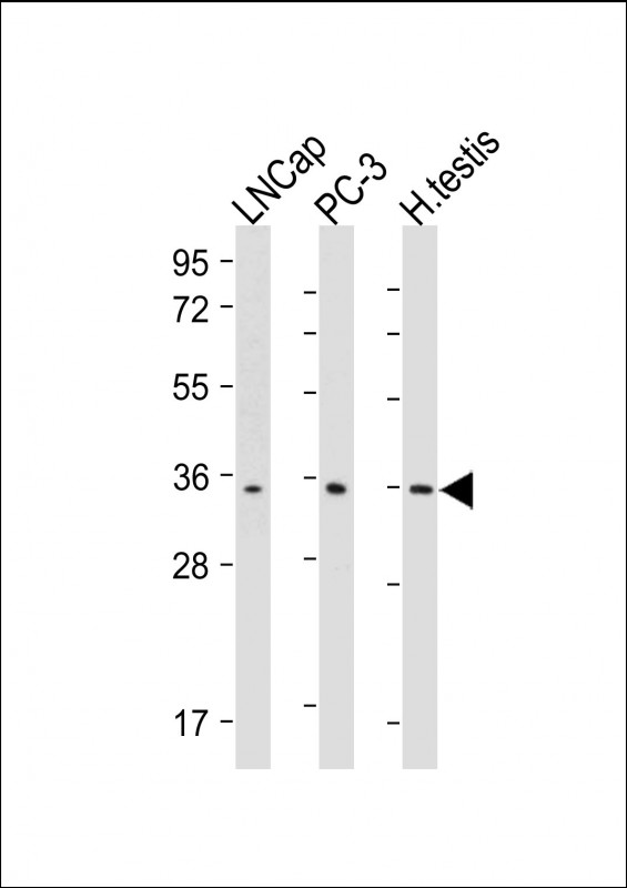
RNF4 Antibody
Purified Mouse Monoclonal Antibody (Mab)
- 产品详情
- 实验流程
- 背景知识
Application
| WB, E |
|---|---|
| Primary Accession | P78317 |
| Reactivity | Human |
| Predicted | Human |
| Host | Mouse |
| Clonality | monoclonal |
| Isotype | IgG1,k |
| Clone Names | 2055CT191.101.249 |
| Calculated MW | 21319 Da |
| Gene ID | 6047 |
|---|---|
| Other Names | E3 ubiquitin-protein ligase RNF4, 6.3.2.-, RING finger protein 4, Small nuclear ring finger protein, Protein SNURF, RNF4, SNURF |
| Target/Specificity | This RNF4 antibody is generated from a mouse immunized with a recombinant protein from the human region of human RNF4. |
| Dilution | WB~~1:2000 E~~Use at an assay dependent concentration. |
| Format | Purified monoclonal antibody supplied in PBS with 0.09% (W/V) sodium azide. This antibody is purified through a protein G column, followed by dialysis against PBS. |
| Storage | Maintain refrigerated at 2-8°C for up to 2 weeks. For long term storage store at -20°C in small aliquots to prevent freeze-thaw cycles. |
| Precautions | RNF4 Antibody is for research use only and not for use in diagnostic or therapeutic procedures. |
| Name | RNF4 {ECO:0000303|PubMed:15815621, ECO:0000312|HGNC:HGNC:10067} |
|---|---|
| Function | E3 ubiquitin-protein ligase which binds polysumoylated chains covalently attached to proteins and mediates 'Lys-6'-, 'Lys-11'-, 'Lys- 48'- and 'Lys-63'-linked polyubiquitination of those substrates and their subsequent targeting to the proteasome for degradation (PubMed:18408734, PubMed:19307308, PubMed:35013556). Regulates the degradation of several proteins including PML and the transcriptional activator PEA3 (PubMed:18408734, PubMed:19307308, PubMed:20943951). Involved in chromosome alignment and spindle assembly, it regulates the kinetochore CENPH-CENPI-CENPK complex by targeting polysumoylated CENPI to proteasomal degradation (PubMed:20212317). Regulates the cellular responses to hypoxia and heat shock through degradation of respectively EPAS1 and PARP1 (PubMed:19779455, PubMed:20026589). Alternatively, it may also bind DNA/nucleosomes and have a more direct role in the regulation of transcription for instance enhancing basal transcription and steroid receptor-mediated transcriptional activation (PubMed:12885770). Catalyzes ubiquitination of sumoylated PARP1 in response to PARP1 trapping to chromatin, leading to PARP1 removal from chromatin by VCP/p97 (PubMed:35013556). |
| Cellular Location | Cytoplasm. Nucleus. Nucleus, PML body |
| Tissue Location | Widely expressed at low levels in many tissues; highly expressed in testis. |
For Research Use Only. Not For Use In Diagnostic Procedures.
Provided below are standard protocols that you may find useful for product applications.
BACKGROUND
E3 ubiquitin-protein ligase which binds polysumoylated chains covalently attached to proteins and mediates 'Lys-6'-, 'Lys-11'-, 'Lys-48'- and 'Lys-63'-linked polyubiquitination of those substrates and their subsequent targeting to the proteasome for degradation. Regulates the degradation of several proteins including PML and the transcriptional activator PEA3. Involved in chromosome alignment and spindle assembly, it regulates the kinetochore CENPH-CENPI-CENPK complex by targeting polysumoylated CENPI to proteasomal degradation. Regulates the cellular responses to hypoxia and heat shock through degradation of respectively EPAS1 and PARP1. Alternatively, it may also bind DNA/nucleosomes and have a more direct role in the regulation of transcription for instance enhancing basal transcription and steroid receptor- mediated transcriptional activation.
REFERENCES
Hadano S.,et al.DNA Res. 5:177-186(1998).
Chiariotti L.,et al.Genomics 47:258-265(1998).
Ota T.,et al.Nat. Genet. 36:40-45(2004).
Hillier L.W.,et al.Nature 434:724-731(2005).
Fedele M.,et al.J. Biol. Chem. 275:7894-7901(2000).
终于等到您。ABCEPTA(百远生物)抗体产品。
点击下方“我要评价 ”按钮提交您的反馈信息,您的反馈和评价是我们最宝贵的财富之一,
我们将在1-3个工作日内处理您的反馈信息。
如有疑问,联系:0512-88856768 tech-china@abcepta.com.


