


 癌症的基本特征包括细胞增殖、血管生成、迁移、凋亡逃避机制和细胞永生等。找到癌症发生过程中这些通路的关键标记物和对应的抗体用于检测至关重要。
 为您推荐一个泛素化位点预测神器——泛素化分析工具,可以为您的蛋白的泛素化位点作出预测和评分。
 细胞自噬受体图形绘图工具为你的蛋白的细胞受体结合位点作出预测和评分,识别结合到自噬通路中的蛋白是非常重要的,便于让我们理解自噬在正常生理、病理过程中的作用,如发育、细胞分化、神经退化性疾病、压力条件下、感染和癌症。
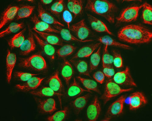
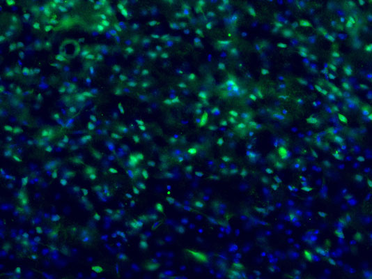
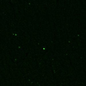
Anti-c-FOS Antibody
Our Anti-c-FOS primary antibody from PhosphoSolutions is mouse monoclonal. It detects bovine, human,

- 产品详情
- 实验流程
- 背景知识
Application
| WB, IHC, ICC |
|---|---|
| Primary Accession | P01100 |
| Host | Mouse |
| Clonality | Monoclonal |
| Isotype | IgG1 |
| Clone Names | 2H2 |
| Calculated MW | 40695 Da |
| Gene ID | 2353 |
|---|---|
| Other Names | Activator protein 1 antibody, AP 1 antibody, C FOS antibody, Cellular oncogene c fos antibody, Cellular oncogene fos antibody, FBJ murine osteosarcoma viral (v fos) oncogene homolog (oncogene FOS) antibody, FBJ murine osteosarcoma viral oncogene homolog antibody, FBJ murine osteosarcoma viral v fos oncogene homolog antibody, FBJ Osteosarcoma Virus antibody, FOS antibody, FOS protein antibody, FOS_HUMAN antibody, G0 G1 switch regulatory protein 7 antibody, G0/G1 switch regulatory protein 7 antibody, G0S7 antibody, Oncogene FOS antibody, p55 antibody, proto oncogene c Fos antibody, Proto oncogene protein c fos antibody, Proto-oncogene c-Fos antibody, fos FBJ murine osteosarcoma viral oncogene homolog antibody |
| Target/Specificity | c-FOS is a member of the FOS transcription factor family which forms dimers with c-JUN to produce the Activator Protein 1 (AP-1) complex which plays a key role in critical cellular processes such as cell proliferation, differentiation and apoptosis (Chiu et al., 1988). c-FOS expression has been demonstrated to be a useful marker of neuronal activation as it is rapidly induced following various stimuli (Hoffman et al., 1993). Additionally, c-FOS has been shown to be overexpressed in a variety of malignant tumor types (Milde-Langosch 2005). |
| Dilution | WB~~1:1000 IHC~~1:100~500 ICC~~N/A |
| Format | Protein G Purified |
| Storage | Maintain refrigerated at 2-8°C for up to 6 months. For long term storage store at -20°C in small aliquots to prevent freeze-thaw cycles. |
| Precautions | Anti-c-FOS Antibody is for research use only and not for use in diagnostic or therapeutic procedures. |
| Shipping | Blue Ice |
For Research Use Only. Not For Use In Diagnostic Procedures.
Provided below are standard protocols that you may find useful for product applications.
BACKGROUND
c-FOS is a member of the FOS transcription factor family which forms dimers with c-JUN to produce the Activator Protein 1 (AP-1) complex which plays a key role in critical cellular processes such as cell proliferation, differentiation and apoptosis (Chiu et al., 1988). c-FOS expression has been demonstrated to be a useful marker of neuronal activation as it is rapidly induced following various stimuli (Hoffman et al., 1993). Additionally, c-FOS has been shown to be overexpressed in a variety of malignant tumor types (Milde-Langosch 2005).
终于等到您。ABCEPTA(百远生物)抗体产品。
点击下方“我要评价 ”按钮提交您的反馈信息,您的反馈和评价是我们最宝贵的财富之一,
我们将在1-3个工作日内处理您的反馈信息。
如有疑问,联系:0512-88856768 tech-china@abcepta.com.


