


 癌症的基本特征包括细胞增殖、血管生成、迁移、凋亡逃避机制和细胞永生等。找到癌症发生过程中这些通路的关键标记物和对应的抗体用于检测至关重要。
 为您推荐一个泛素化位点预测神器——泛素化分析工具,可以为您的蛋白的泛素化位点作出预测和评分。
 细胞自噬受体图形绘图工具为你的蛋白的细胞受体结合位点作出预测和评分,识别结合到自噬通路中的蛋白是非常重要的,便于让我们理解自噬在正常生理、病理过程中的作用,如发育、细胞分化、神经退化性疾病、压力条件下、感染和癌症。
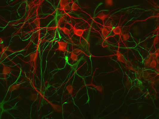
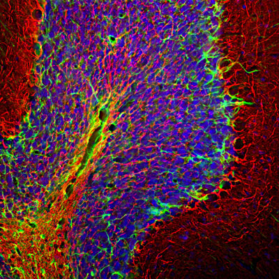
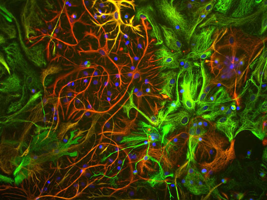
Anti-Glial Fibrillary Acidic Protein (GFAP) Antibody
Our Anti-Glial Fibrillary Acidic Protein (GFAP) primary antibody from PhosphoSolutions is rabbit pol
- 产品详情
- 实验流程
- 背景知识
Application
| WB, IHC, ICC |
|---|---|
| Primary Accession | P14136 |
| Host | Rabbit |
| Clonality | Polyclonal |
| Isotype | IgG |
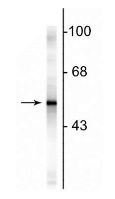
| Calculated MW | 49880 Da |
| Gene ID | 2670 |
|---|---|
| Other Names | wu:fb34h11 antibody, ALXDRD antibody, cb345 antibody, etID36982.3 antibody, FLJ42474 antibody, FLJ45472 antibody, GFAP antibody, GFAP_HUMAN antibody, gfapl antibody, Glial fibrillary acidic protein antibody, Intermediate filament protein antibody, wu:fk42c12 antibody, xx:af506734 antibody, zgc:110485 antibody |
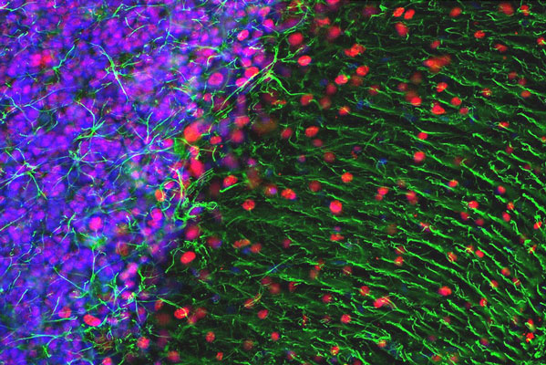
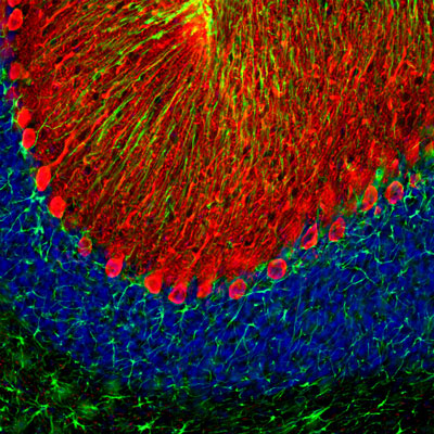
| Target/Specificity | Glial Fibrillary Acidic Protein (GFAP) was discovered by Amico Bignami and co-workers as a major fibrous protein of multiple sclerosis plaques (1). It was subsequently found to be a member of the 10nm or intermediate filament (IF) family, specifically the IF family Class III, which also includes peripherin, desmin and vimentin. GFAP is strongly and specifically expressed in astrocytes and certain other astroglia in the CNS, in satellite cells, peripheral ganglia, and in non-myelinating Schwann cells in peripheral nerves. In many damage and disease states GFAP expression is heavily upregulated in astrocytes. In addition, neural stem cells frequently strongly express GFAP. Point mutations in the protein coding region of the GFAP gene lead to Alexander disease which is characterized by the presence of abnormal astrocytes containing GFAP protein aggregates known as Rosenthal fibers (2). |
| Dilution | WB~~1:1000 IHC~~1:100~500 ICC~~N/A |
| Format | Neat Serum |
| Storage | Maintain refrigerated at 2-8°C for up to 6 months. For long term storage store at -20°C in small aliquots to prevent freeze-thaw cycles. |
| Precautions | Anti-Glial Fibrillary Acidic Protein (GFAP) Antibody is for research use only and not for use in diagnostic or therapeutic procedures. |
| Shipping | Blue Ice |
For Research Use Only. Not For Use In Diagnostic Procedures.
Provided below are standard protocols that you may find useful for product applications.
BACKGROUND
Glial Fibrillary Acidic Protein (GFAP) was discovered by Amico Bignami and co-workers as a major fibrous protein of multiple sclerosis plaques (1). It was subsequently found to be a member of the 10nm or intermediate filament (IF) family, specifically the IF family Class III, which also includes peripherin, desmin and vimentin. GFAP is strongly and specifically expressed in astrocytes and certain other astroglia in the CNS, in satellite cells, peripheral ganglia, and in non-myelinating Schwann cells in peripheral nerves. In many damage and disease states GFAP expression is heavily upregulated in astrocytes. In addition, neural stem cells frequently strongly express GFAP. Point mutations in the protein coding region of the GFAP gene lead to Alexander disease which is characterized by the presence of abnormal astrocytes containing GFAP protein aggregates known as Rosenthal fibers (2).
终于等到您。ABCEPTA(百远生物)抗体产品。
点击下方“我要评价 ”按钮提交您的反馈信息,您的反馈和评价是我们最宝贵的财富之一,
我们将在1-3个工作日内处理您的反馈信息。
如有疑问,联系:0512-88856768 tech-china@abcepta.com.


