


 癌症的基本特征包括细胞增殖、血管生成、迁移、凋亡逃避机制和细胞永生等。找到癌症发生过程中这些通路的关键标记物和对应的抗体用于检测至关重要。
 为您推荐一个泛素化位点预测神器——泛素化分析工具,可以为您的蛋白的泛素化位点作出预测和评分。
 细胞自噬受体图形绘图工具为你的蛋白的细胞受体结合位点作出预测和评分,识别结合到自噬通路中的蛋白是非常重要的,便于让我们理解自噬在正常生理、病理过程中的作用,如发育、细胞分化、神经退化性疾病、压力条件下、感染和癌症。
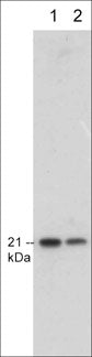
Anti-p21/CIP1/WAF1 Antibody
- 产品详情
- 实验流程
- 背景知识
Application
| WB, IHC, ICC, IP |
|---|---|
| Primary Accession | P38936 |
| Host | Mouse |
| Clonality | Mouse Monoclonal |
| Isotype | IgG2a |
| Clone Names | M513 |
| Calculated MW | 18119 Da |
| Gene ID | 1026 |
|---|---|
| Other Names | Cyclin-dependent kinase inhibitor 1 CDK-interacting protein 1 Melanoma differentiation-associated protein 6 MDA-6 p21 CDKN1A CAP20, CDKN1, CIP1, MDA6, PIC1, SDI1, WAF1 |
| Dilution | WB~~1:1000 IHC~~1:100~500 ICC~~N/A IP~~N/A |
| Storage | Maintain refrigerated at 2-8°C for up to 6 months. For long term storage store at -20°C in small aliquots to prevent freeze-thaw cycles. |
| Precautions | Anti-p21/CIP1/WAF1 Antibody is for research use only and not for use in diagnostic or therapeutic procedures. |
| Shipping | Blue Ice |
For Research Use Only. Not For Use In Diagnostic Procedures.
Provided below are standard protocols that you may find useful for product applications.
BACKGROUND
The tumor suppressor protein p21/CIP1/WAF1 acts as an inhibitor of cell cycle progression. It functions in stoichiometric relationships forming heterotrimeric complexes with cyclins and cyclin-dependent kinases. In association with CDK2 complexes, it serves to inhibit kinase activity and block progression through G1/S. However, p21 may also enhance assembly and activity in complexes of CDK4 or CDK6 and cyclin D. The carboxy-terminal region of p21 is sufficient to bind and inhibit PCNA, a subunit of DNA polymerase, and may coordinate DNA replication with cell cycle progression. Upon UV damage or during cell cycle stages when cdc2/cyclin B or CDK2/cyclin A are active, p53 is phosphorylated and upregulates p21 transcription via a p53-responsive element. Protein levels of p21 are downregulated through ubiquitination and proteasomal degradation.
终于等到您。ABCEPTA(百远生物)抗体产品。
点击下方“我要评价 ”按钮提交您的反馈信息,您的反馈和评价是我们最宝贵的财富之一,
我们将在1-3个工作日内处理您的反馈信息。
如有疑问,联系:0512-88856768 tech-china@abcepta.com.


