


 癌症的基本特征包括细胞增殖、血管生成、迁移、凋亡逃避机制和细胞永生等。找到癌症发生过程中这些通路的关键标记物和对应的抗体用于检测至关重要。
 为您推荐一个泛素化位点预测神器——泛素化分析工具,可以为您的蛋白的泛素化位点作出预测和评分。
 细胞自噬受体图形绘图工具为你的蛋白的细胞受体结合位点作出预测和评分,识别结合到自噬通路中的蛋白是非常重要的,便于让我们理解自噬在正常生理、病理过程中的作用,如发育、细胞分化、神经退化性疾病、压力条件下、感染和癌症。
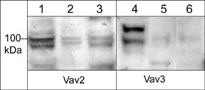
Anti-Vav3 Antibody
- 产品详情
- 实验流程
- 背景知识
Application
| WB |
|---|---|
| Primary Accession | Q9UKW4 |
| Host | Rabbit |
| Clonality | Rabbit Polyclonal |
| Isotype | IgG |
| Calculated MW | 97776 Da |
| Gene ID | 10451 |
|---|---|
| Other Names | VAV3, Guanine nucleotide exchange factor VAV3 |
| Dilution | WB~~1:1000 |
| Storage | Maintain refrigerated at 2-8°C for up to 6 months. For long term storage store at -20°C in small aliquots to prevent freeze-thaw cycles. |
| Precautions | Anti-Vav3 Antibody is for research use only and not for use in diagnostic or therapeutic procedures. |
| Shipping | Blue Ice |
For Research Use Only. Not For Use In Diagnostic Procedures.
Provided below are standard protocols that you may find useful for product applications.
BACKGROUND
The Vav family of Rho-guanine nucleotide exchange factors, Vav1, Vav2, and Vav3, have central roles in transducing signals from cell surface receptors, such as integrin, growth factor and immune cell receptors to the cytoskeleton. This role includes receptor-mediated changes in the actin cytoskeleton and cell motility. Vav1 expression is normally restricted to hematopoietic cells, while Vav2 and Vav3 are more widely expressed. All three Vav isoforms have been shown to be abnormally expressed in several types of cancer. Vavs are composed of multiple domains, including a Dbl homology domain, a calponin homology domain, an acidic amino acid region, a pleckstrin homology domain, a cysteine-rich domain, and SH3 and SH2 domains. Vav activity is regulated by the phosphorylation status of several conserved tyrosine residues in the acidic region (In Vav2: Tyr-142, Tyr-159, and Tyr-172). These tyrosine residues are able to participate in autoinhibitory interactions with the Dbl homology domain and this interaction is prevented after phosphorylation of these sites leading to activation of Vav GEF activity.
终于等到您。ABCEPTA(百远生物)抗体产品。
点击下方“我要评价 ”按钮提交您的反馈信息,您的反馈和评价是我们最宝贵的财富之一,
我们将在1-3个工作日内处理您的反馈信息。
如有疑问,联系:0512-88856768 tech-china@abcepta.com.


