


 癌症的基本特征包括细胞增殖、血管生成、迁移、凋亡逃避机制和细胞永生等。找到癌症发生过程中这些通路的关键标记物和对应的抗体用于检测至关重要。
 为您推荐一个泛素化位点预测神器——泛素化分析工具,可以为您的蛋白的泛素化位点作出预测和评分。
 细胞自噬受体图形绘图工具为你的蛋白的细胞受体结合位点作出预测和评分,识别结合到自噬通路中的蛋白是非常重要的,便于让我们理解自噬在正常生理、病理过程中的作用,如发育、细胞分化、神经退化性疾病、压力条件下、感染和癌症。
Calcyclin Antibody
Purified Mouse Monoclonal Antibody
- 产品详情
- 实验流程
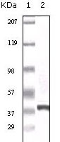
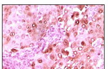
Application
| WB, IHC, E |
|---|---|
| Primary Accession | Q9HB71 |
| Reactivity | Human |
| Host | Mouse |
| Clonality | Monoclonal |
| Clone Names | 7D11 |
| Isotype | IgG1 |
| Calculated MW | 26210 Da |
| Description | Calcyclin encoded by this gene is a member of the S100 family of proteins containing 2 EF-hand calcium-binding motifs. S100 proteins are localized in the cytoplasm and/or nucleus of a wide range of cells, and involved in the regulation of a number of cellular processes such as cell cycle progression and differentiation. S100 genes include at least 13 members which are located as a cluster on chromosome 1q21. This protein may function in stimulation of Ca2+-dependent insulin release, stimulation of prolactin secretion, and exocytosis. Chromosomal rearrangements and altered expression of this gene have been implicated in melanoma. |
| Immunogen | Purified recombinant fragment of calcyclin expressed in E. Coli. |
| Formulation | Purified antibody in PBS containing 0.03% sodium azide. |
| Gene ID | 27101 |
|---|---|
| Other Names | Calcyclin-binding protein, CacyBP, hCacyBP, S100A6-binding protein, Siah-interacting protein, CACYBP, S100A6BP, SIP |
| Dilution | WB~~1/500 - 1/2000 IHC~~1/200 - 1/1000 E~~N/A |
| Storage | Maintain refrigerated at 2-8°C for up to 6 months. For long term storage store at -20°C in small aliquots to prevent freeze-thaw cycles. |
| Precautions | Calcyclin Antibody is for research use only and not for use in diagnostic or therapeutic procedures. |
| Name | CACYBP |
|---|---|
| Synonyms | S100A6BP, SIP |
| Function | May be involved in calcium-dependent ubiquitination and subsequent proteasomal degradation of target proteins. Probably serves as a molecular bridge in ubiquitin E3 complexes. Participates in the ubiquitin-mediated degradation of beta-catenin (CTNNB1). |
| Cellular Location | Nucleus. Cytoplasm. Note=Cytoplasmic at low calcium concentrations. In neuroblastoma cells, after a retinoic acid (RA) induction and calcium increase, it localizes in both the nucleus and cytoplasm. The nuclear fraction may be phosphorylated |
Research Areas
For Research Use Only. Not For Use In Diagnostic Procedures.
Application Protocols
Provided below are standard protocols that you may find useful for product applications.
REFERENCES
1. Bean C.et.al J Mol Biol. 2005 Jun 3;349(2):349-66. 2. HWang R.et.al J Biol Chem. 2004 May 14;279(20):21239-47.
终于等到您。ABCEPTA(百远生物)抗体产品。
点击下方“我要评价 ”按钮提交您的反馈信息,您的反馈和评价是我们最宝贵的财富之一,
我们将在1-3个工作日内处理您的反馈信息。
如有疑问,联系:0512-88856768 tech-china@abcepta.com.


