


 癌症的基本特征包括细胞增殖、血管生成、迁移、凋亡逃避机制和细胞永生等。找到癌症发生过程中这些通路的关键标记物和对应的抗体用于检测至关重要。
 为您推荐一个泛素化位点预测神器——泛素化分析工具,可以为您的蛋白的泛素化位点作出预测和评分。
 细胞自噬受体图形绘图工具为你的蛋白的细胞受体结合位点作出预测和评分,识别结合到自噬通路中的蛋白是非常重要的,便于让我们理解自噬在正常生理、病理过程中的作用,如发育、细胞分化、神经退化性疾病、压力条件下、感染和癌症。
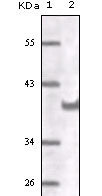
CD31 Antibody
Purified Mouse Monoclonal Antibody
- 产品详情
- 实验流程
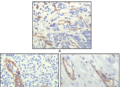
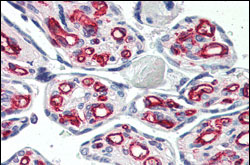
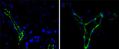
Application
| WB, IHC, ICC, E |
|---|---|
| Primary Accession | P16284 |
| Reactivity | Human |
| Host | Mouse |
| Clonality | Monoclonal |
| Clone Names | 2F7B2 |
| Isotype | IgG1 |
| Calculated MW | 82522 Da |
| Description | CD31, also known as platelet endothelial cell adhesion molecule 1 (PECAM1), is a type I integral membrane glycoprotein and a member of the immunoglobulin superfamily of cell surface receptors.It is constitutively expressed on the surface of endothelial cells, and concentrated at the junction between them. The antibody reacts with the murine form of the Platelet-Endothelial Cell Adhesion Molecule. The reactivity of the antibody is restricted to the isoform of the molecule that is selectively expressed by endothelial cells.The antigen is predominantly present at the lateral borders of endothelial cells as described for human PECAM-1.It is also weakly expressed on many peripheral lymphoid cells and platelets.CD31 has been used to measure angiogenesis in association with tumor recurrence. Other studies have also indicated that CD31 and CD34 can be used as markers for myeloid progenitor cells and recognize different subsets of myeloid leukemia infiltrates (granular sarcomas). |
| Immunogen | Purified recombinant fragment of human CD31 expressed in E. Coli. |
| Formulation | Ascitic fluid containing 0.03% sodium azide. |
| Gene ID | 5175 |
|---|---|
| Other Names | Platelet endothelial cell adhesion molecule, PECAM-1, EndoCAM, GPIIA', PECA1, CD31, PECAM1 |
| Dilution | WB~~1/500 - 1/2000 IHC~~1/500 - 1/2000 ICC~~N/A E~~N/A |
| Storage | Maintain refrigerated at 2-8°C for up to 6 months. For long term storage store at -20°C in small aliquots to prevent freeze-thaw cycles. |
| Precautions | CD31 Antibody is for research use only and not for use in diagnostic or therapeutic procedures. |
| Name | PECAM1 |
|---|---|
| Function | Cell adhesion molecule which is required for leukocyte transendothelial migration (TEM) under most inflammatory conditions (PubMed:17580308, PubMed:19342684). Tyr-690 plays a critical role in TEM and is required for efficient trafficking of PECAM1 to and from the lateral border recycling compartment (LBRC) and is also essential for the LBRC membrane to be targeted around migrating leukocytes (PubMed:19342684). Trans-homophilic interaction may play a role in endothelial cell-cell adhesion via cell junctions (PubMed:27958302). Heterophilic interaction with CD177 plays a role in transendothelial migration of neutrophils (PubMed:17580308). Homophilic ligation of PECAM1 prevents macrophage-mediated phagocytosis of neighboring viable leukocytes by transmitting a detachment signal (PubMed:12110892). Promotes macrophage-mediated phagocytosis of apoptotic leukocytes by tethering them to the phagocytic cells; PECAM1-mediated detachment signal appears to be disabled in apoptotic leukocytes (PubMed:12110892). Modulates bradykinin receptor BDKRB2 activation (PubMed:18672896). Regulates bradykinin- and hyperosmotic shock-induced ERK1/2 activation in endothelial cells (PubMed:18672896). Induces susceptibility to atherosclerosis (By similarity). |
| Cellular Location | Cell membrane; Single-pass type I membrane protein. Note=Cell surface expression on neutrophils is down-regulated upon fMLP or CXCL8/IL8- mediated stimulation. [Isoform Delta15]: Cell junction. Note=Localizes to the lateral border recycling compartment (LBRC) and recycles from the LBRC to the junction in resting endothelial cells |
| Tissue Location | Expressed on platelets and leukocytes and is primarily concentrated at the borders between endothelial cells (PubMed:18388311, PubMed:21464369). Expressed in human umbilical vein endothelial cells (HUVECs) (at protein level) (PubMed:17580308, PubMed:19342684). Expressed on neutrophils (at protein level) (PubMed:17580308). Isoform Long predominates in all tissues examined (PubMed:12433657). Isoform Delta12 is detected only in trachea (PubMed:12433657). Isoform Delta14-15 is only detected in lung (PubMed:12433657). Isoform Delta14 is detected in all tissues examined with the strongest expression in heart (PubMed:12433657). Isoform Delta15 is expressed in brain, testis, ovary, cell surface of platelets, human umbilical vein endothelial cells (HUVECs), Jurkat T- cell leukemia, human erythroleukemia (HEL) and U-937 histiocytic lymphoma cell lines (at protein level) (PubMed:12433657, PubMed:18388311). |
Research Areas
For Research Use Only. Not For Use In Diagnostic Procedures.
Application Protocols
Provided below are standard protocols that you may find useful for product applications.
REFERENCES
1. Mayr U et al. Circ Res 98:412-20 (2006). 2. Bingle L et al. Br J Cancer 94:101-7 (2006). 3. Wynne F et al. Reproduction 131:721-32 (2006).
终于等到您。ABCEPTA(百远生物)抗体产品。
点击下方“我要评价 ”按钮提交您的反馈信息,您的反馈和评价是我们最宝贵的财富之一,
我们将在1-3个工作日内处理您的反馈信息。
如有疑问,联系:0512-88856768 tech-china@abcepta.com.
¥ 1,500.00
Cat# AO1104a


