


 癌症的基本特征包括细胞增殖、血管生成、迁移、凋亡逃避机制和细胞永生等。找到癌症发生过程中这些通路的关键标记物和对应的抗体用于检测至关重要。
 为您推荐一个泛素化位点预测神器——泛素化分析工具,可以为您的蛋白的泛素化位点作出预测和评分。
 细胞自噬受体图形绘图工具为你的蛋白的细胞受体结合位点作出预测和评分,识别结合到自噬通路中的蛋白是非常重要的,便于让我们理解自噬在正常生理、病理过程中的作用,如发育、细胞分化、神经退化性疾病、压力条件下、感染和癌症。
TNF-alpha Antibody
Purified Mouse Monoclonal Antibody
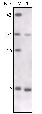
- 产品详情
- 实验流程
Application 
| WB, E |
|---|---|
| Primary Accession | P01375 |
| Reactivity | Human |
| Host | Mouse |
| Clonality | Monoclonal |
| Clone Names | 2A9B9 |
| Isotype | IgG1 |
| Calculated MW | 25644 Da |
| Description | TNF-alpha (tumor necrosis factor alpha) is an important cytokine produced by numerous cell types including neutrophils, activated lymphocytes, macrophages and NK cells. It plays a critical role in inflammatory responses and in apoptosis. TNF-alpha is believed to mediate pathogenic shock and tissue injury associated with endotoxemia. TNF-alpha exists as a multimer of two, three, or five non covalently linked units, but shows a single 17 kDa band following SDS PAGE under non reducing conditions. Although it has little effect on many cultured normal human cells, TNF-alpha appears to be directly toxic to vascular endothelial cells. Other actions of TNF-alpha include stimulating growth of human fibroblasts and other cell lines, activating polymorphonuclear neutrophils and osteoclasts, and induction of interleukin 1, prostaglandin E2 and collagenase production. TNF-alpha is currently being evaluated in treatment of certain cancers and AIDS Related Complex. |
| Immunogen | Recombinant Human TNF-alpha (BioSource company, Cat.No. PHC3013) |
| Formulation | Purified antibody in PBS containing 0.03% sodium azide. |
| Gene ID | 7124 |
|---|---|
| Other Names | Tumor necrosis factor, Cachectin, TNF-alpha, Tumor necrosis factor ligand superfamily member 2, TNF-a, Tumor necrosis factor, membrane form, N-terminal fragment, NTF, Intracellular domain 1, ICD1, Intracellular domain 2, ICD2, C-domain 1, C-domain 2, Tumor necrosis factor, soluble form, TNF, TNFA, TNFSF2 |
| Dilution | WB~~1/500 - 1/2000 E~~N/A |
| Storage | Maintain refrigerated at 2-8°C for up to 6 months. For long term storage store at -20°C in small aliquots to prevent freeze-thaw cycles. |
| Precautions | TNF-alpha Antibody is for research use only and not for use in diagnostic or therapeutic procedures. |
For Research Use Only. Not For Use In Diagnostic Procedures.
| Name | TNF |
|---|---|
| Synonyms | TNFA, TNFSF2 |
| Function | Cytokine that binds to TNFRSF1A/TNFR1 and TNFRSF1B/TNFBR. It is mainly secreted by macrophages and can induce cell death of certain tumor cell lines. It is potent pyrogen causing fever by direct action or by stimulation of interleukin-1 secretion and is implicated in the induction of cachexia, Under certain conditions it can stimulate cell proliferation and induce cell differentiation. Impairs regulatory T- cells (Treg) function in individuals with rheumatoid arthritis via FOXP3 dephosphorylation. Up-regulates the expression of protein phosphatase 1 (PP1), which dephosphorylates the key 'Ser-418' residue of FOXP3, thereby inactivating FOXP3 and rendering Treg cells functionally defective (PubMed:23396208). Key mediator of cell death in the anticancer action of BCG-stimulated neutrophils in combination with DIABLO/SMAC mimetic in the RT4v6 bladder cancer cell line (PubMed:16829952, PubMed:22517918, PubMed:23396208). Induces insulin resistance in adipocytes via inhibition of insulin-induced IRS1 tyrosine phosphorylation and insulin-induced glucose uptake. Induces GKAP42 protein degradation in adipocytes which is partially responsible for TNF-induced insulin resistance (By similarity). Plays a role in angiogenesis by inducing VEGF production synergistically with IL1B and IL6 (PubMed:12794819). Promotes osteoclastogenesis and therefore mediates bone resorption (By similarity). |
| Cellular Location | Cell membrane; Single-pass type II membrane protein [Tumor necrosis factor, soluble form]: Secreted [C-domain 2]: Secreted. |
Research Areas
Application Protocols
Provided below are standard protocols that you may find useful for product applications.
REFERENCES
1. Knowlton KU. Yajima T. J Am Coll Cardiol. 2004,Sep 15, 44(6):1298-300. 2. Reynolds JL. Ignatowski TA. Gallant S.et al. Brain Res. 2004,Oct 8, 1023(1):112-20. 3. Dulak J. Tomala K. Loboda A. et al. Life Sci. 2004,Oct 8, 75(21):2573-86.

终于等到您。ABCEPTA(百远生物)抗体产品。
点击下方“我要评价 ”按钮提交您的反馈信息,您的反馈和评价是我们最宝贵的财富之一,
我们将在1-3个工作日内处理您的反馈信息。
如有疑问,联系:0512-88856768 tech-china@abcepta.com.
¥ 1,250.00
Cat# AO1134a


