


 癌症的基本特征包括细胞增殖、血管生成、迁移、凋亡逃避机制和细胞永生等。找到癌症发生过程中这些通路的关键标记物和对应的抗体用于检测至关重要。
 为您推荐一个泛素化位点预测神器——泛素化分析工具,可以为您的蛋白的泛素化位点作出预测和评分。
 细胞自噬受体图形绘图工具为你的蛋白的细胞受体结合位点作出预测和评分,识别结合到自噬通路中的蛋白是非常重要的,便于让我们理解自噬在正常生理、病理过程中的作用,如发育、细胞分化、神经退化性疾病、压力条件下、感染和癌症。
CRYAB Antibody
Purified Mouse Monoclonal Antibody
- 产品详情
- 实验流程
Application
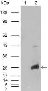
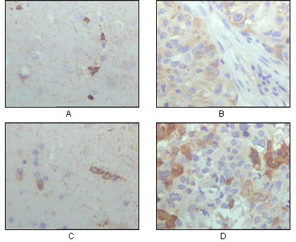
| WB, IHC, E |
|---|---|
| Primary Accession | P02511 |
| Reactivity | Human |
| Host | Mouse |
| Clonality | Monoclonal |
| Clone Names | 1D11C6E6 |
| Isotype | IgG2a |
| Calculated MW | 20159 Da |
| Description | Crystallin, alpha B. Crystallins are separated into two classes: taxon-specific, or enzyme, and ubiquitous. The latter class constitutes the major proteins of vertebrate eye lens and maintains the transparency and refractive index of the lens. Since lens central fiber cells lose their nuclei during development, these crystallins are made and then retained throughout life, making them extremely stable proteins. Mammalian lens crystallins are divided into alpha, beta, and gamma families; beta and gamma crystallins are also considered as a superfamily. Alpha and beta families are further divided into acidic and basic groups. Seven protein regions exist in crystallins: four homologous motifs, a connecting peptide, and N- and C-terminal extensions. Alpha crystallins are composed of two gene products: alpha-A and alpha-B, for acidic and basic, respectively. Alpha crystallins can be induced by heat shock and are members of the small heat shock protein (sHSP also known as the HSP20) family. They act as molecular chaperones although they do not renature proteins and release them in the fashion of a true chaperone; instead they hold them in large soluble aggregates. Post-translational modifications decrease the ability to chaperone. These heterogeneous aggregates consist of 30-40 subunits; the alpha-A and alpha-B subunits have a 3:1 ratio, respectively. Two additional functions of alpha crystallins are an autokinase activity and participation in the intracellular architecture. Alpha-A and alpha-B gene products are differentially expressed; alpha-A is preferentially restricted to the lens and alpha-B is expressed widely in many tissues and organs. Elevated expression of alpha-B crystallin occurs in many neurological diseases; a missense mutation cosegregated in a family with a desmin-related myopathy. |
| Immunogen | Purified recombinant fragment of CRYAB (aa1-175) expressed in E. Coli. |
| Formulation | Ascitic fluid containing 0.03% sodium azide. |
| Gene ID | 1410 |
|---|---|
| Other Names | Alpha-crystallin B chain, Alpha(B)-crystallin, Heat shock protein beta-5, HspB5, Renal carcinoma antigen NY-REN-27, Rosenthal fiber component, CRYAB, CRYA2 |
| Dilution | WB~~1/500 - 1/2000 IHC~~1/500 - 1/2000 E~~N/A |
| Storage | Maintain refrigerated at 2-8°C for up to 6 months. For long term storage store at -20°C in small aliquots to prevent freeze-thaw cycles. |
| Precautions | CRYAB Antibody is for research use only and not for use in diagnostic or therapeutic procedures. |
| Name | CRYAB (HGNC:2389) |
|---|---|
| Synonyms | CRYA2, HSPB5 |
| Function | May contribute to the transparency and refractive index of the lens. Has chaperone-like activity, preventing aggregation of various proteins under a wide range of stress conditions. In lens epithelial cells, stabilizes the ATP6V1A protein, preventing its degradation by the proteasome (By similarity). |
| Cellular Location | Cytoplasm. Nucleus Secreted. Lysosome {ECO:0000250|UniProtKB:P23927}. Note=Translocates to the nucleus during heat shock and resides in sub-nuclear structures known as SC35 speckles or nuclear splicing speckles (PubMed:19464326). Localizes at the Z- bands and the intercalated disk in cardiomyocytes (PubMed:28493373) Can be secreted; the secretion is dependent on protein unfolding and facilitated by the cargo receptor TMED10; it results in protein translocation from the cytoplasm into the ERGIC (endoplasmic reticulum- Golgi intermediate compartment) followed by vesicle entry and secretion (PubMed:32272059). |
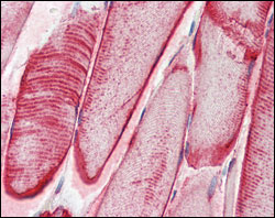
| Tissue Location | Lens as well as other tissues (PubMed:2387586, PubMed:838078). Expressed in myocardial tissue (PubMed:28493373) |
Research Areas
For Research Use Only. Not For Use In Diagnostic Procedures.
Application Protocols
Provided below are standard protocols that you may find useful for product applications.
REFERENCES
1. Cell. 2007 Aug 10;130(3):427-39. 2. Biochemistry. 2006 Nov 21;45(46):13847-54. 3. J Mol Biol. 2007 Sep 14;372(2):470-84.
终于等到您。ABCEPTA(百远生物)抗体产品。
点击下方“我要评价 ”按钮提交您的反馈信息,您的反馈和评价是我们最宝贵的财富之一,
我们将在1-3个工作日内处理您的反馈信息。
如有疑问,联系:0512-88856768 tech-china@abcepta.com.
¥ 1,500.00
Cat# AO1152a


