


 癌症的基本特征包括细胞增殖、血管生成、迁移、凋亡逃避机制和细胞永生等。找到癌症发生过程中这些通路的关键标记物和对应的抗体用于检测至关重要。
 为您推荐一个泛素化位点预测神器——泛素化分析工具,可以为您的蛋白的泛素化位点作出预测和评分。
 细胞自噬受体图形绘图工具为你的蛋白的细胞受体结合位点作出预测和评分,识别结合到自噬通路中的蛋白是非常重要的,便于让我们理解自噬在正常生理、病理过程中的作用,如发育、细胞分化、神经退化性疾病、压力条件下、感染和癌症。
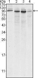
Calnexin Antibody
Purified Mouse Monoclonal Antibody
- 产品详情
- 实验流程
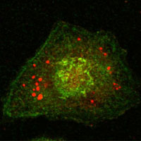
Application
| WB, ICC, E |
|---|---|
| Primary Accession | P27824 |
| Reactivity | Human |
| Host | Mouse |
| Clonality | Monoclonal |
| Clone Names | 3H4A7 |
| Isotype | IgG2b |
| Calculated MW | 67568 Da |
| Description | This gene encodes a member of the calnexin family of molecular chaperones. The encoded protein is a calcium-binding, endoplasmic reticulum (ER)-associated protein that interacts transiently with newly synthesized N-linked glycoproteins, facilitating protein folding and assembly. It may also play a central role in the quality control of protein folding by retaining incorrectly folded protein subunits within the ER for degradation. Alternatively spliced transcript variants encoding the same protein have been described. |
| Immunogen | Synthetic peptide corresponding to aa (CEAAEERPWLWVVYILTVAL) of human Calnexin, conjugated to KLH. |
| Formulation | Ascitic fluid containing 0.03% sodium azide. |
| Gene ID | 821 |
|---|---|
| Other Names | Calnexin, IP90, Major histocompatibility complex class I antigen-binding protein p88, p90, CANX |
| Dilution | WB~~1/500 - 1/2000 ICC~~N/A E~~N/A |
| Storage | Maintain refrigerated at 2-8°C for up to 6 months. For long term storage store at -20°C in small aliquots to prevent freeze-thaw cycles. |
| Precautions | Calnexin Antibody is for research use only and not for use in diagnostic or therapeutic procedures. |
| Name | CANX |
|---|---|
| Function | Calcium-binding protein that interacts with newly synthesized monoglucosylated glycoproteins in the endoplasmic reticulum. It may act in assisting protein assembly and/or in the retention within the ER of unassembled protein subunits. It seems to play a major role in the quality control apparatus of the ER by the retention of incorrectly folded proteins. Associated with partial T-cell antigen receptor complexes that escape the ER of immature thymocytes, it may function as a signaling complex regulating thymocyte maturation. Additionally it may play a role in receptor-mediated endocytosis at the synapse. |
| Cellular Location | Endoplasmic reticulum membrane; Single-pass type I membrane protein. Mitochondrion membrane {ECO:0000250|UniProtKB:P24643}; Single-pass type I membrane protein. Melanosome membrane; Single-pass type I membrane protein. Note=Identified by mass spectrometry in melanosome fractions from stage I to stage IV (PubMed:12643545, PubMed:17081065). The palmitoylated form preferentially localizes to the perinuclear rough ER (PubMed:22314232) Localizes to endoplasmic reticulum mitochondria-associated membrane (MAMs) that connect the endoplasmic reticulum and the mitochondria (By similarity). {ECO:0000250|UniProtKB:P24643, ECO:0000269|PubMed:12643545, ECO:0000269|PubMed:17081065, ECO:0000269|PubMed:22314232} |
Research Areas
For Research Use Only. Not For Use In Diagnostic Procedures.
Application Protocols
Provided below are standard protocols that you may find useful for product applications.
REFERENCES
1. Science. 2003 Feb 28;299(5611):1394-7. 2. Exp Cell Res. 2004 Mar 10;294(1):244-53. 3. Science. 2004 Apr 23;304(5670):600-2.
终于等到您。ABCEPTA(百远生物)抗体产品。
点击下方“我要评价 ”按钮提交您的反馈信息,您的反馈和评价是我们最宝贵的财富之一,
我们将在1-3个工作日内处理您的反馈信息。
如有疑问,联系:0512-88856768 tech-china@abcepta.com.
¥ 1,500.00
Cat# AO1158a


